- Home
- Users & Science
- Scientific Documentation
- ESRF Highlights
- ESRF Highlights 2003
- X-ray Absorption and Magnetic Scattering
- Resonant Magnetic X-ray Scattering in Co-doped CeFe2
Resonant Magnetic X-ray Scattering in Co-doped CeFe2
The magnetic ground state of CeFe2 has historically been controversial. Although the ground states of the heavy rare-earth (RE) laves phases with Fe (formula REFe2) are well understood, that of CeFe2 is considerably more complex as a consequence of the hybridisation of the anomalous f electrons in Ce, with the 3d states of iron [1].
Resonant Magnetic X-ray scattering (RMXS) experiments performed on a Co-doped single crystal of CeFe2 (grown at Ames Laboratory, Iowa State University, Ames, Iowa USA) allowed us to re-examine this challenging problem.
 |
|
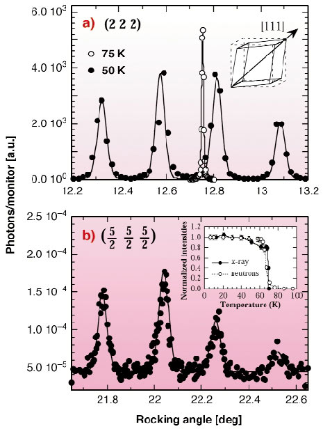
Fig. 123: a) Splitting of the crystallographic Bragg (222) reflection due to the rhombohedral distortion; b) Magnetic reflection (5/2 5/2 5/2) split in the corresponding rhombohedral domains. |
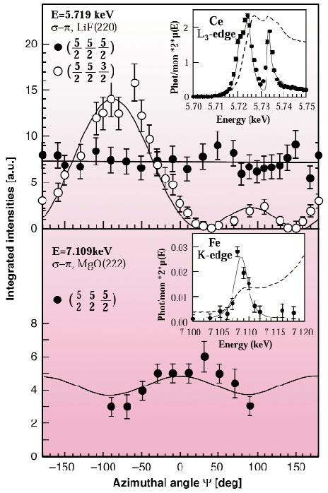
The experiments were carried out at beamline ID20 on a sample of CeFe2 doped with 7% Co, at which concentration the antiferromagnetic ground state is stabilised below 69 K. Between this temperature and the Curie temperature of 210 K the material is a "nominal" ferromagnet, as is the pure compound. Below the discontinuous antiferromagnetic transition, a rhombohedral distortion splits the Bragg peaks along the <111> directions into four crystallographic domains. Exploiting the high Q-resolution available on ID20, it was possible to isolate the contribution of a single magnetic domain, as shown in Figure 123. By tuning the incident photon energy to the Ce L3 and at the Fe K edges on performing linear polarisation analysis of the scattered photons, it was possible through the resonant X-ray scattering process to probe the magnetically-polarised 4f and the 3d electronic states respectively. Since these processes are also sensitive to the magnetic moment directions, by rotating the sample around the scattering vector (indicate azimuthal scans), we can determine the moment direction of both Ce and Fe magnetic sublattices. The results indicate that the Ce moments to be parallel to the propagation vector [1/2 1/2 1/2] within each magnetic domain. The excellent fit of such a simple model to the experimental points in Figure 124 (top panel) leaves little doubt as to this assignment. The difficulty is that all previous neutron-diffraction results appear inconsistent with this interpretation [2]. In addition, Mössbauer spectroscopy (sensitive to the Fe atoms) was inconsistent with a simple collinear arrangement of the Fe moments [3]. Indeed, the azimuthal scans at the Fe K-edge (Figure 124, bottom panel) shows a modulation in the azimuthal dependence, indicating that not all the Fe moments can lie along the [111] direction. Unfortunately, however, the enhancement at the Fe K edge is so weak that a definite assignment of the Fe moment orientation in the AF state cannot be obtained with RMXS.
 |
|
Fig. 124: Azimuthal dependence of RMXS intensities of the magnetic reflections (5/2 5/2 5/2) and (5/2 5/2 3/2) at the Ce L3 edge (upper panel) and at the Fe K-edge (lower panel). The insets show the resonant enhancement around the Ce and Fe absorption edges. |
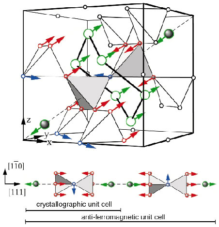
A neutron experiment was then designed at the D10 instrument, Institute Laue Langevin, using a single crystal and gathering a larger data set than previously collected with polycrystalline samples. The analysis of this data set (330 reflections measured, reducing to 72 inequivalent data points) showed unambiguously that the Fe moments are noncollinear. The resulting configuration is shown in Figure 125. Indeed the molecular field provided by the Fe moments has its major component along the [111] direction in Figure 125; thus forcing the much smaller Ce moments to be along this direction (in agreement with the synchrotron experiments as shown in Figure 124). However, in addition, there is a noncollinear component with the smaller Fe moment of 1.1 µB.
 |
|
Fig. 125: Magnetic structure of the antiferromagnetic state obtained combining neutron and RMXS scattering results. The smaller "blue" Fe moments are indeed dynamic in nature, both in the antiferromagnetic ground state and in the ferromagnetic state. |
The resulting model can be used to rationalise many of the unusual and conflicting experimental results reported for this material in literature, and can explain the dynamical properties of the magnetic ground state found in pure CeFe2 [4].
References
[1] O. Eriksson et al., Phys. Rev. Lett. 60, 2523 (1988).
[2] S. J. Kennedy and B. Coles, J. Phys. Cond. Mat. 2, 1213 (1990).
[3] J-P. Sanchez et al., Hyperfine Interact. 133, 5 (2001).
[4] L. Paolasini et al, Phys. Rev. Lett. 90, 057201 (2003); Phys. Rev. B 58, 12117 (1998).
Authors
L. Paolasini (a), B. Ouladdiaf (b), N. Bernhoeft (c), JP. Sanchez (c), P. Vulliet (c), G. H. Lander (d), and P. Canfield (e)
(a) ESRF
(b) ILL
(c) CEA-Grenoble (France)
(d) European Commission, JRC, Karlsruhe (Germany)
(e) Iowa State University (USA)


