- Home
- Users & Science
- Scientific Documentation
- ESRF Highlights
- ESRF Highlights 2005
- X-ray Imaging and Optics
- New Irradiation Geometry for Microbeam Radiation Therapy (MRT)
New Irradiation Geometry for Microbeam Radiation Therapy (MRT)
Microbeam Radiation Therapy (MRT) has the potential to treat infantile brain tumours when other kinds of radiotherapy would be excessively toxic to the developing normal brain [1, 2]. MRT uses extraordinarily high doses of X-rays but the brain provides unusual resistance to MRT’s radioneurotoxicity, presumably by the migration of endothelial cells from “valleys” into “peaks”, i.e., into directly irradiated microslices of tissues. A novel irradiation geometry that results in an even more tolerable valley dose for the normal tissue and a decreased peak-to-valley dose ratio (PVDR) in the tumour area by applying an innovative cross-firing technique has been studied. It consists of orthogonally crossfiring two arrays of parallel, nonintersecting, mutually interspersed microbeams that produces tumouricidal doses with small PVDRs where the arrays meet and tolerable radiation doses to normal tissues between the microbeams proximal and distal to the tumour in the paths of the arrays.
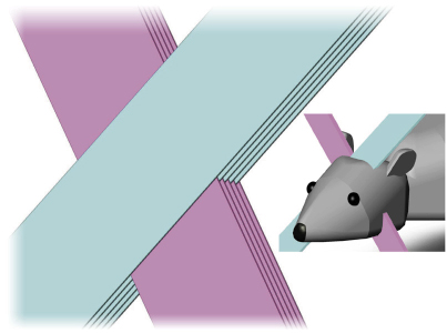
A possible interlaced crossfiring geometry is shown in Figure 144 with the microbeams at +45 degrees and –45 degrees. Before the second exposure, the target is rotated 900 around the axis and then displaced by 105 µm in the plane perpendicular to the direction of propagation of the microbeams, i.e. half the distance between two microbeams. The latter displacement was to interlace rather than intersect the orthogonally-propagated microbeam planes.
 |
|
Fig. 144: interlaced cross-firing technique. |
The use of microbeams as a potential alternative in radiotherapy is attractive because of the destruction of the vascularisation of the tumour and also because of the high tolerance of these microbeams by normal tissue. This dose-volume effect might therefore be used in the future in radiotherapy where crucial tissue must be spared and selectively different sensitivities of the vasculature between tumour and the surrounding tissue can be exploited. We can further enhance this effect by crossing the microbeams to selectively distribute the valley dose in the tumour to a radiotoxic value and leave the valley dose in the healthy tissue at acceptable values.
 |
|
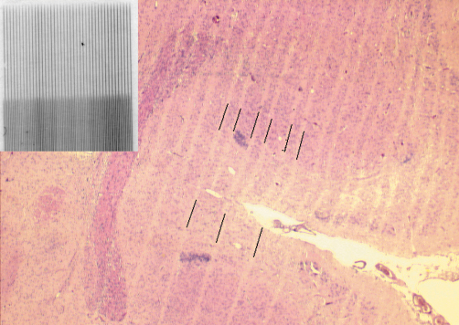
Fig. 145: the interlaced feature of our cross-fired microbeams is still preserved inside the brain of the irradiated specimen; the inserted picture showing the mechanically precise irradiation on a Gafchromic film. |
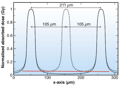
The mechanical feasibility of this technique was demonstrated using Gafchromic films, but in order to answer the question of whether the interlaced feature of our cross-fired microbeams is still preserved inside the brain of the irradiated specimen, the horizontal histology cuts of such irradiated rats are shown in Figure 145. The parallelism of the microbeams is well preserved inside the tissue, despite the microscopic movements due to the pulsation of the heart beat and a non-ridged target like the brain. According to our Monte Carlo calculations we profit from an increase of the valley dose by about a factor of 3 (Figure 146).
 |
|
Fig. 146: Schematic illustration of PVDRs and the decrease of valley dose if the second irradiation plane is centred with its peak situated between the peaks of the first irradiation. |
There are several methods for increasing the therapeutic index of MRT; first by limiting the cross-fired zone more closely to the actual tumour area. This will be the case when an adequate imaging technique is available and the densely irradiated volume can be reduced to the actual tumour size. Additionally, a better understanding between the physical and biological correlation of parameters like microbeam planar width and centre-to-centre distances is needed to optimise the parameters which can then be selected more specifically for the healthy tissue area and the tumour region. Moreover, one can expect even better results if we move to larger volumes to be irradiated, since in this case the ratio between the total volume of the brain, the irradiated volume and the more densely crossfired tumour volume become more favourable. Additionally, we can alleviate the limitation of tumour dose, and thereby the limitation of therapeutic efficiency due to skin dose thresholds.
Conventional radiation therapy is hampered by the acceptable threshold dose of the healthy tissue. By using microbeams in an interlaced irradiation geometry, we can take advantage of healthy tissues extraordinarily good tolerance of microbeams and hence deliver a critical level of radiation only to the vicinity of the tumour. The result is a nearly homogenous distribution of high dose that is limited to the tumour area.
References
[1] J. A. Laissue, G. Geiser, P. O. Spanne, F. A. Dilmanian, J-O. Gebbers, M. Geiser, X. Y. Wu, M. S. Makar, P. L. Micca, M. M. Nawrocky, D. D. Joel, and D. N. Slatkin, Int. J. Cancer 78, 654-660 (1998).
[2) J. A. Laissue, N. Lyubimova, H.-P. Wagner, D. W. Archer, D. N. Slatkin, M. Di Michiel, C. Nemoz, M. Renier, E. Brauer, P. O. Spanne, J.-O. Gebbers, K. Dixon, and H. Blattmann, Proceedings of SPIE Vol.3770, 38-45 (1999).
Principal Publication and Authors
E. Brauer-Krisch (a), H. Requardt (a), P. Régnard (a), S. Corde (a), E. Siegbahn (a), G. LeDuc (a), T. Brochard (a), H. Blattmann (b), J. Laissue (c), A. Bravin (a), Phys. Med. Biol. 50, 3103-3111 (2005).
(a) ESRF
(b) Niederwiesstrasse 13C, Untersiggenthal (Switzerland)
(c) Institute of Pathology, University of Bern (Switzerland)


