- Home
- Users & Science
- Scientific Documentation
- ESRF Highlights
- ESRF Highlights 2005
- Soft Condensed Matter
- Hard X-ray Nanoprobe Based on Refractive X-ray Lenses
Hard X-ray Nanoprobe Based on Refractive X-ray Lenses
Hard X-ray scanning microscopy allows one to perform X-ray analytical techniques, such as X-ray fluorescence analysis, absorption spectroscopy, or diffraction, with high spatial resolution. In this way, element distributions, the chemical state of an element, or the local (crystalline) structure of a heterogeneous specimen can be determined. In combination with tomographic techniques, local information from the inside of a sample can be obtained without destructive sample preparation. This is beneficial to many fields of science, such as chemistry, materials, earth, environmental, and biomedical science. These hard X-ray scanning microscopy techniques greatly benefit from the recent advances in X-ray optics generating ever smaller and more intense nanobeams. Today, a variety of X-ray optics is capable of generating beams with a lateral beam size in the range of 100 nm and below. Among these are nanofocusing lenses (NFLs) [1,2], on the basis of which we are currently developing a hard X-ray nanoprobe station at beamline ID13. This hard X-ray scanning microscope can be operated in transmission, fluorescence, and diffraction mode. Tomographic scanning modes will allow one to obtain elemental and structural information from inside a specimen.
At the heart of the microscope are nanofocusing refractive X-ray lenses that image the synchrotron radiation source onto the sample position in a strongly reducing geometry. Their key strength is their short focal length that lies in the centimetre range. In this way, they can generate a sub-100 nm beam even at short source to experiment distances and do not require particularly long beamlines. In addition, the diffraction limit of these optics typically lies well below 50 nm. In their current design (cf. Figure 72a), these planar lenses generate a line focus. The latest generation of these optics is made of Si using lithographic processes optimised for 4''-wafers. In this way, the fabrication parameters are well controlled, reducing shape errors to a minimum. Several lenses are placed side by side on the same wafer. Figure 72a shows a scanning electron micrograph of part of such a set of lenses. Each of these lenses has a slightly different focal length for a given X-ray energy. In the microscope, the X-ray nanobeam is generated by two crossed nanofocusing lenses that are aligned along a common optical axis as shown in Figure 72b. The common aperture of the lens system is defined by a guard aperture.
 |
|
Fig. 72: (a) Scanning electron micrograph of nanofocusing lenses. A single lens and a nanofocusing lens are outlined by dark shaded areas. (b) Nanoprobe setup: the X-ray beam is focused onto the sample by two crossed nanofocusing lenses. |
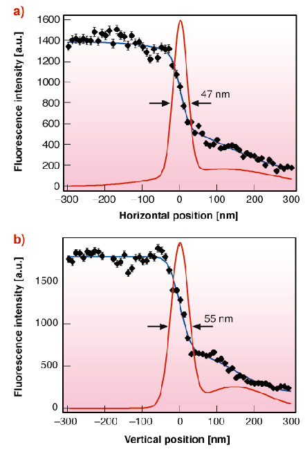
During the first commissioning phase of the scanning microscope at beamline ID13 the nanobeam was characterised. It was set up at 47 m from the in-vacuum undulator source. Using a pair of nanofocusing lenses with a focal length of fh, = 10.7 mm and fv = 19.4 mm for horizontal and vertical focusing, respectively, a hard X-ray beam (E = 21 keV) with a lateral extension of 47 x 55 nm2 was generated (Figure 73). The flux in this beam is 1.7 x 10⁸ ph/s at a ring current of 200 mA, resulting in a gain in flux density of more than 2 x 10⁴. In the future, this beam will be used for fluorescence tomography and nanodiffraction.
 |
|
Fig. 73: (a) Horizontal and (b) vertical beam profile determined by fluorescence knife-edge scans. (Reprinted with permission from [1].) |
The main advantage of this setup is that extremely small foci can be generated even at short beamlines. Using the available fabrication technology for a new lens scheme (adiabatically focusing lenses [3]), beams as small as 20 nm are conceivable in the current geometry. In addition, using other lens materials, such as boron or diamond, the flux and focus size can also be significantly improved. Besides scanning microscopy other techniques may profit from a small focus, such as diffraction with coherent X-rays and X-ray photon and fluorescence correlation spectroscopy.
References
[2] C.G. Schroer, M. Kuhlmann, U.T. Hunger, T.F. Günzler, O. Kurapova, S. Feste, F. Frehse, B. Lengeler, M. Drakopoulos, A. Somogyi, A.S. Simionovici, A. Snigirev, I. Snigireva, C. Schug, W.H. Schröder, Appl. Phys. Lett. 82, 1485 (2003).
[3] C. G. Schroer and B. Lengeler, Phys. Rev. Lett. 94, 054802 (2005).
Principal Publication and Authors
[1] C.G. Schroer (a), O. Kurapova (b), J. Patommel (b), P. Boye (b), J. Feldkamp (b), B. Lengeler (b), M. Burghammer (c), C. Riekel (c), L. Vincze (d), A. van der Hart (e), M. Küchler (f), Appl. Phys. Lett. 87(12), 124103 (2005).
(a) HASYLAB at DESY, Hamburg (Germany)
(b) II. Physikalisches Institut, Aachen University (Germany)
(c) ESRF
(d) Department of Analytical Chemistry, Ghent University (Belgium)
(e) ISG, Forschungszentrum Jülich (Germany)
(f) Fraunhofer IZM, Dept. Microdevices and Equipment (Germany)


