- Home
- Users & Science
- Scientific Documentation
- ESRF Highlights
- ESRF Highlights 2007
- X-ray Imaging and Optics
X-ray Imaging and Optics
Introduction
X-ray imaging techniques are being used more and more frequently in modern synchrotron radiation facilities. The feature common to all of these techniques is that they apply to inhomogeneous samples, where it is important to measure “locally” a given property such as density, composition, chemical state or distortion. These techniques take advantage of most of the photon-matter interactions: absorption, wavefront modification, diffraction, scattering, photoemission, etc. An increasing part of the experimental results obtained at the ESRF can now be considered as “X-ray images”, i.e. maps over the sample of the “local” value of a physical quantity. In this case “local” does not mean atomic level (whereas in some cases atomic information can be extracted from the images) but corresponds to the very important 10–3-10–8 m range, where many biological and materials science phenomena occur.
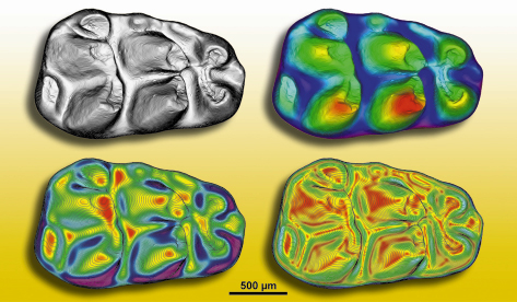
These microscopy techniques apply to a wide variety of topics. Indeed, this chapter shows contributions in biological and biomedical applications (iron storage within dopamine neurovesicles, the microvascular network in brain, improved three dimensional imaging of breast tissues), materials science (3D real-time observation of solidification in binary alloys, optical luminescence microscopy in GaN), and also applications to Cultural Heritage (study of micrometric pigment in Grunewald’s paintings, occurrence in early homo sapiens of modern human development). Paleontology is an emerging scientific area at the ESRF and other synchrotron radiation facilities: the number of paleontological proposals has increased from zero to 20% of the proposals for microtomography over the last five years. As an example, Figure 120 shows various quantitative maps computed from X-ray synchrotron microtomographic data, of the external morphology of a molar of an ancestor of rats and mice, which allow a good comparison of the morphological details of several similar fossils, and an identification of the mastication directions of this animal.
 |
|
Fig. 120: Synchrotron radiation microtomographic maps of the external morphology of the first lower molar of Progonomys cathalai (late Miocene, from Montredon, South of France). Top left corner: 3D rendering colour topographic map. Top right corner: colour topographic map with contour lines. Bottom left corner: colour slope map with contour lines. Bottom right corner: colour angularity map with contour lines. Depth between two consecutive contour lines: 12.5 µm (From Lazzari, Tafforeau, Aguilar and Michaux, Paleobiology, 2008). |
The importance of synchrotron radiation-based X-ray imaging techniques is reflected by the considerable effort of ESRF users and staff in pushing the limits of the techniques in terms of resolution and domains of applications, and in starting new techniques. This effort is exemplified in the present chapter by contributions on scanning X-ray microscopy to study microbes involvement in geochemical cycles and near field X-ray speckles.
A substantial part of the ESRF Science and Technology Programme 2008-2017 relies on improved imaging techniques. The spatial resolution is heading towards the nanometre scale, and high temporal resolution will increasingly allow chemical and industrial processes to be followed in situ. Developments exploiting the partial coherence of synchrotron X-ray beams for phase contrast imaging or coherent diffraction imaging will continue. This requires a combined effort, high throughput imaging being strongly dependent on detector improvements and computing for the reconstruction of holographic images from a set of radiographs, and the iterative determination of the phase of the scattering amplitude in coherent diffraction imaging. Developments in all of these areas are going to occur in the very near future.
J. Baruchel


