- Home
- Users & Science
- Scientific Documentation
- ESRF Highlights
- ESRF Highlights 2009
- X-ray imaging
- Tumour functional mapping using synchrotron radiation computed tomography
Tumour functional mapping using synchrotron radiation computed tomography
Dynamic contrast enhanced medical imaging (DCE-MI), using MRI (magnetic resonance imaging) or CT (computed tomography), is a promising technique for the detection, characterisation and monitoring of many lesions such as solid tumours. Its use has recently been extended to in vivo assessment of the microcirculatory function of the tissues. An intravenous bolus of contrast agent is followed as a function of time in tissues through local variations of grey levels in pixels or in regions of pixels. The evolution of the contrast agent concentration depends on microcirculatory characteristics which are assessed by using pharmacokinetic modelling. Several phenomena can hamper microcirculatory assessments and, in practice, the variability in data acquisition techniques and data analysis is so important between research centres and/or hospitals that results cannot be easily compared. Thus, objective quantification of microcirculatory parameters by this technique could appear limited.
To test the potential of DCE-MI in terms of microcirculatory parameter quantification, we analysed images acquired from a controlled DCE-MI protocol in order to minimise potential artefacts or bias that could hamper the results. We tested whether it was possible to characterise different structural elements of C6 rat brain tumours by their differing microcirculatory behaviour. The acquisition was provided in stereotactic conditions using the monochromatic synchrotron radiation CT (SRCT) technique available at ID17, the Biomedical Beamline. Compared to other techniques, SRCT offers a high signal to noise ratio and does not suffer from beam hardening. In particular, the technique allows the measurement of absolute rather than relative values. The analysis was provided by selecting the more relevant pharmacokinetic model among several compartmental models, for the set of selected anatomic regions. The model selection was guided by using quality criteria [1] that tested the consistency between original data and modelling information provided by each pharmacokinetic model. This a posteriori analysis avoids any potential misinterpretation of results and helps in evaluating the reliability of the final parametric results.
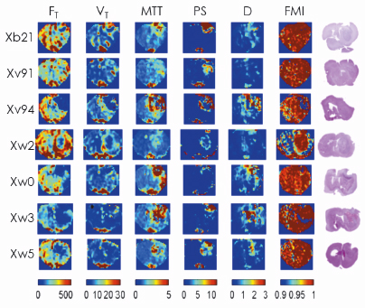
DCE-SRCT sequences were analysed using purpose-built software (PhysioD3D). The software provided parametric maps of tumours and surrounding brain tissues, each map corresponding to different microcirculatory parameters: blood flow, blood volume, mean transit time, capillary permeability index and artery to tissue delay (Figure 127). It also provided specific maps for quality control. Structure regions were selected in the original sequences, guided by the parametric maps and corresponding post mortem histological slices. Finally microcirculatory parameters in the defined regions were compared.
 |
|
Fig. 127: Examples of parametric maps of c6 glioma-bearing rat brains displayed alongside the corresponding histological slices, for 7 rats. From left to right: blood flow (FT), blood volume (VT), mean transit time (MTT), permeability (PS), artery to tissue delay (D), quality criteria (FMI, in red for optimal quality) and histological slices. |
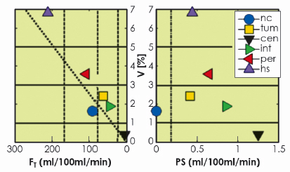
Remarkably, the regional organisation of the tumour zones, visible on the hematoxylin-eosin-stained slices, were reproduced on a parametric map for which high quality criteria were found (Figure 127). Quantitative results in the selected regions (Figure 128) indicated that each type of tumour tissue is characterised by abnormal capillary permeability, due to a damaged blood-brain barrier, and by lower intravascular transit time (V/FT). Quantitative results also indicated that the internal structures of the tumour could be characterised by their specific blood flow and blood volume values, increasing in the following order: centre, intermediate, periphery and hot spots.
 |
|
Fig. 128: Microcirculation characteristics of tumour and healthy tissues for blood flow (FT), blood volume (V) and permeability (PS). The different regions: healthy caudate nucleus (nc), global tumour (tum), and tumour structures: centre (cen), intermediate zone (int), periphery (per) and hot spots (hs) have different averaged microcirculatory parameters (markers) corresponding to significant microcirculatory behaviour when markers are separated by a line. |
The consistency between DCE-SRCT and histological maps such as the ability of DCE-SRCT to significantly differentiate microcirculatory behaviour between tumour elements could position this technique as a reference. In particular, the assessment of absolute concentrations allows comparison between centres and estimation of calibration factors.
References
[1] D. Balvay, F. Frouin, G. Calmon and C.A. Cuenod, Magn. Reson. Med. 54, 868-877 (2005).
Principal publication and authors
D. Balvay (a), I. Troprès (b), R. Billet (c), A. Joubert (c), M. Péoc’h (c), C.A. Cuenod (a) and G. Le Duc (c), Radiology 250, 692-702 (2009).
(a) LRI, PARCC, Inserm 970 – Service Radiologie HEGP, Paris (France)
(b) IFR1, Université Joseph Fourier – Unité IRM 3T, CHU Grenoble (France)
(c) ESRF


