- Home
- Users & Science
- Scientific Documentation
- ESRF Highlights
- ESRF Highlights 2009
- X-ray imaging
- Sub-micrometre holotomography of multiphase metals
Sub-micrometre holotomography of multiphase metals
The microstructural characterisation of multiphase materials by conventional 2D metallography can be insufficient if, for instance, connectivity between phases exists, or the orientation of constituents varies throughout the volume. In this case, 3D characterisation tools are needed. Synchrotron microtomography is a powerful technique capable of revealing the architecture of materials. Furthermore, the coherence of the beam can be exploited by applying quantitative phase-contrast tomography or holotomography [1] to image components with similar attenuation. The spatial resolution achievable by parallel beam synchrotron microtomography is about 1 µm. This can be improved using magnifying optics [2]. So far this has been achieved for samples with diameters < 100 µm using relatively low energies. 3D imaging of engineering alloys requires high energies and representative sample sizes to correlate the properties with the microstructure achieved by the processing method.
Magnified synchrotron holotomography using a Kirkpatrick-Baez (KB) optics system [3] was carried out at the nanoimaging end-station ID22NI for Al- and Ti-alloy samples of 0.4 mm diameter. The focal point with a size of 80 nm (H) by 130 nm (V) and medium monochromaticity (![]() E/E = 2x10⁻²) was produced by a set of multilayer coated crossed bent mirrors. Energies of 17.5 keV and 29 keV as well as effective pixel sizes of 60 nm and 51 nm were used for the Al-alloy and Ti-alloys, respectively. Phase retrieval for holotomography was achieved from recordings at four focal-point-to-sample distances.
E/E = 2x10⁻²) was produced by a set of multilayer coated crossed bent mirrors. Energies of 17.5 keV and 29 keV as well as effective pixel sizes of 60 nm and 51 nm were used for the Al-alloy and Ti-alloys, respectively. Phase retrieval for holotomography was achieved from recordings at four focal-point-to-sample distances.
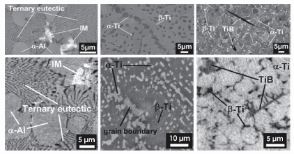
Examples of three light alloys are presented. Their microstructures are shown in the scanning electron micrographs (SEM) at the top of Figure 136:
- AlMg7Si4 alloy: the microstructure exhibits a fine ternary eutectic formed by Al, Si and Mg2Si with lamellar structures >150 nm. Fe-rich intermetallics appear within the primary
 -Al.
-Al. - Ti-10V-2Fe-3Al alloy (Ti1023): the alloy contains ~30% of hcp a phase distributed within bcc ß grains not observable by absorption contrast.
- Ti-6Al-4V (Ti64) alloy reinforced with 5 vol% TiB needles (Ti64/TiB/5w): the Ti64 alloy matrix consists of
 grains separated by narrow ß zones and TiB needles.
Holotomography is well adapted to these materials as there is low absorption contrast between the present phases.
 |
|
Fig. 136 :From left to right: The materials are AlMg7Si4 alloy, Ti1023 alloy and Ti64/TiB/5w. Upper images are scanning electron micrographs; lower images are portions of slices of holotomographic reconstructions (magnified holotomography using KB optics). |
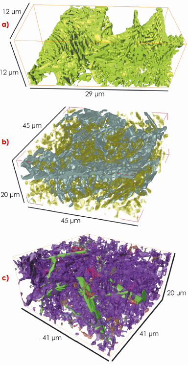
Portions of the reconstructed holotomography slices are shown at the bottom of Figure 136. The grey level scales linearly with the electron density of the constituents revealing all the phases observed in the SEM micrographs. Volume views of the minority phases are shown in Figure 137.
- AlMg7Si4: the ternary eutectic with interconnected Si-Mg2Si structures as thin as ~ 180 nm (green) are identified. The intermetallic particles and a-Al are made transparent in Figure 137a.
- Ti1023:
 grains > 200 nm are identified. In Figure 137b, an interconnected structure of a particles remained from an incomplete break-up of the lamellar structure during pre-forging (blue), while secondary
  grains (green) are embedded in the ß phase.
 - Ti64/TiB/5w: the
 and ß phases in the Ti64 matrix are identified down to 200 nm. In Figure 137c, TiB needles (green) are connected to irregularly shaped ß grains (other colours), forming an interpenetrating structure within the
  phase.
 |
|
Fig. 137 : Rendered volumes of the materials: a) interconnected Si-Mg2Si structure (green) in AlMg7Si4; b) larger a particles remaining from pre-forging (blue) and individual secondary |
In summary, magnified synchrotron holotomography using KB optics was used to characterise the 3D-architecture of Al- and Ti-alloy samples of ~0.4 mm diameter with energies of 17.5 and 29 keV, respectively. Using the nano-focusing KB-system for such a high energy (29 keV) is reported for the first time. This experimental setup combining synchrotron radiation, KB optics and holotomography opens a new level of resolution (≥ 180 nm) for non-destructive 3D imaging of representative samples of multiphase metals.
References
[1] P. Cloetens, W. Ludwig, J, Baruchel, D. Van Dyck, J. Van Landuyt, J.P. Guigay and M. Schlenker, Appl. Phys. Lett. 75, 2912-2914 (1999).
[2] P. J. Withers, Materials Today 10, 26-34 (2007).
[3] R. Mokso, P. Cloetens, E. Maire, W. Ludwig and J.Y. Buffiere, Appl. Phys. Lett. 90, 144104 (2007).
Principal publication and authors
G. Requena (a), P. Cloetens (b), W. Altendorfer (a), C. Poletti (a), D. Tolnai (a,c), F. Warchomicka (a) and H. P. Degischer (a), Scripta Materialia 61, 760-763 (2009).
(a) Institute of Materials Science and Technology, Vienna University of Technology, Vienna (Austria)
(b) ESRF
(c) Eötvös Loránd University, Department of Materials Physics, Budapest (Hungary)


