- Home
- Users & Science
- Scientific Documentation
- ESRF Highlights
- ESRF Highlights 2011
- X-ray imaging
- Two-dimensional grating interferometer
Two-dimensional grating interferometer
Phase-contrast imaging with ultra high sensitivity is among the unique applications of modern X-ray sources. With X-ray grating interferometry, a particularly powerful phase-contrast method, details with tiny mass density differences have been successfully imaged with hard X-rays and at spatial resolutions of a few micrometres [1]. X-ray grating interferometry also provides ‘dark-field’ images that reveal the presence of features such as cracks and nanopores smaller than the spatial resolution of the imaging system. Phase and dark-field signals complement, and significantly improve, the conventional absorption information. Grating-based imaging has therefore been attracting interest from many fields, including biology, medicine and materials science.
The standard one-dimensional X-ray grating interferometer (GIFM) consists of two line gratings: the phase grating G1 and the absorption grating G2, both placed between the sample and the detector. The interference pattern generated by G1, which contains information on the optical properties of the sample, is analysed with G2 in order to retrieve the differential phase and dark-field image signals in the direction perpendicular to the grating lines. In the past two years, a GIFM of this kind has been made available to external users at beamline ID19. Recent results include the visualisation of the architecture and cells of the human brain [1] and a study on the orientation and scattering strength of dentinal tubules in teeth [2].
A collaboration between the Paul Scherrer Institute (Switzerland), the Karlsruhe Institute of Technology (Germany), Synchrotron Soleil (France) and the ESRF has significantly improved this imaging technique and broadened its application areas by developing a two-dimensional (2D) GIFM. This 2D GIFM gives access to differential phase and dark-field signals in multiple directions of the image plane.
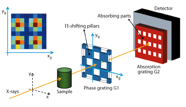
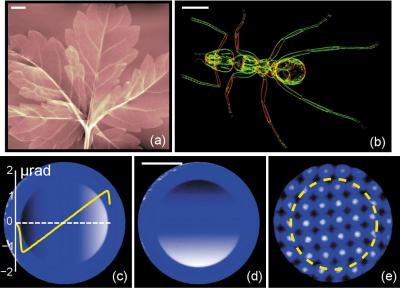
Figure 143 shows a schematic representation of the novel device. The phase grating G1 has a checkerboard structure [3] with silicon pillars that shift the X-ray wave phase by 180°. The analyser grating G2 has strongly absorbing structures made of gold. The image signals are retrieved by a raster scan of G1 in the lateral directions xg and yg (“phase stepping”). The intensity recorded in each detector pixel during the phase-stepping scan can be approximated by a 2D sinusoid (inset in Figure 143). The position and amplitude of the sinusoid in x and y give, respectively, differential phase and dark-field signals along the two directions. Examples of phase and dark-field radiographs of test samples obtained with the 2D GIFM are shown in Figure 144a and b.
Due to the availability of contrast in x and y, the information content and the quality of the final images provided by the 2D GIFM are much higher than with a 1D device.
The 2D GIFM measures refraction angles with a precision down to 10 nrad, which makes it particularly interesting for the at-wavelength characterisation of X-ray optics. The potential of the 2D GIFM in metrology has recently been demonstrated on parabolic refractive lenses (Figure 144c and d) [4].
For applications which require that information is extracted from a single shot, moiré imaging based on the 2D GIFM can be used, instead of the phase-stepping technique. In this case, however, the spatial resolution is limited by the period of the moiré pattern (Figure 144e). Single-shot 2D moiré imaging is particularly promising for fast multimodal tomography and wave-front diagnostics at X-ray free-electron lasers.
Principal publication and authors
I. Zanette (a), T. Weitkamp (a,b), T. Donath (c), S. Rutishauser (c) and C. David (c), Phys. Rev. Lett. 105, 24102 (2010).
(a) ESRF
(b) Synchrotron Soleil, Gif-sur-Yvette (France)
(c) Laboratory for Micro- and Nanotechnology, Paul Scherrer Institute, Villigen (Switzerland)
References
[1] G. Schulz, T. Weitkamp, I. Zanette, F. Pfeiffer, F. Beckmann, C. David, S. Rutishauser, E. Reznikova and B. Müller, J. R. Soc. Interface 7, 1665-1676 (2010).
[2] T.H. Jensen, M. Bech, I. Zanette, T. Weitkamp, C. David, H. Deyhle, S. Rutishauser, E. Reznikova, J. Mohr, R. Feidenhans'l and F. Pfeiffer, Phys. Rev. B 82, 214103 (2010).
[3] J. Primot and N. Guérineau, Appl. Opt. 39, 5715-5720 (2000).
[4] S. Rutishauser, I. Zanette, T. Weitkamp, T. Donath and C. David, Appl. Phys. Lett. 99, 221104 (2011).


