- Home
- Users & Science
- Scientific Documentation
- ESRF Highlights
- ESRF Highlights 2012
- X-ray imaging
- Towards reduced radiation dose for 3-D breast cancer scans
Towards reduced radiation dose for 3-D breast cancer scans
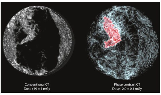
Early detection of breast cancer contributes to an improved prognosis and results in reduced disease mortality. The breast cancer screening technique used today in clinics is “dual-view digital mammography”. Its limitation is that it only provides two images of the breast, which can explain why up to 20% of breast tumours are not detectable on mammograms. Mammograms can also sometimes appear abnormal, when no breast cancers are actually present. Computed tomography (CT) allows a precise 3-D visualisation of the human body but it cannot be routinely applied in breast cancer diagnosis because of the high radiosensitivity of the breast, which significantly reduces the benefit-risk ratio. The potential induction of tumours due to the exam itself is considered too high. Recognising these limitations, we have developed a way to produce three-dimensional X-ray images of the breast at a radiation dose that is lower than the 2D radiographies used in clinics. Phase contrast X-ray tomography at 60 keV of a whole human breast has been performed at ID17. Images were reconstructed applying the so-called “equally sloped tomography” (EST) algorithm using 512 projections to produce 3-D images of the organ at a resolution higher than that used in clinical CT (pixel size of 92 μm vs more than 200 μm for clinical CT scanners) and at a lower dose than that of dual view mammography (which is typically about 3 mGy). The new method allows a clear and precise identification of a malignant cancer. According to a blind evaluation by five experienced radiologists, this method can reduce the radiation dose and acquisition time by ~74% relative to conventional phase-contrast X-ray tomography, while maintaining high resolution and contrast. Despite the significant reduction in the number of projections used (corresponding to an important saving in dose and acquisition time), radiologists ranked the generated images as having the highest sharpness, contrast, and overall image quality compared to 3-D images of breast tissue created through other standard methods (based on the filtered back projection algorithm (FBP)) (Figure 56).
CT scans for early detection of breast cancer may now become possible thanks to the combination of three ingredients: high energy X-rays, phase contrast imaging and the use of a sophisticated novel mathematical algorithm, the EST, to reconstruct the CT images from X-ray data. Tissues are more transparent to high energy X-rays and therefore less dose is deposited (a factor of 6 in radiation dose reduction). The phase contrast imaging technique may allow contrast to be observed due to the phase modulation of X-rays, even if the amplitude (i.e. absorption) modulation is weak or absent, with a dose to the tissues similar or even reduced compared to conventional absorption radiography. Finally, the EST method is a Fourier-based iterative method that iterates back and forth between real and Fourier space using the pseudo-polar fast Fourier transform. At each iteration, physical constraints, including the sample boundary and the non-negativity of the sample structure, are enforced in real space, while the Fourier transform of the measured projections are imposed in Fourier space. As a result, the EST needs 4 times less radiation than FBP to produce images of the same quality (Figure 57).
 |
|
Fig. 57: Image quality comparison of the phase contrast CT images of the whole breast (9 cm diameter), reconstructed using the conventional (Hamming filtered) FBP algorithm or the EST. a) A 92-μm-thick sagittal slice of the FBP reconstruction using 2000 projections, b) EST using 512 projections or c) EST 200. The yellow ellipse indicates a tumour region. FBP 2000 and EST 512 have the highest image quality, EST 200 (with 1/10 of the dose necessary to reconstruct the FBP2000) still shows a high contrast. |
It was therefore demonstrated that diagnostic CT images can be produced with a spatial resolution 2-3 times higher than present hospital scanners, but with a radiation dose that is about 25 times lower. This new technique can open the doors to the clinical use of CT in mamography, which would be a powerful tool in the fight against breast cancer.
Principal publication and authors
Y. Zhao (a), E. Brun (b,c), P. Coan (c,d), Z. Huang (a), A. Sztrókay (d), P. C. Diemoz (c), S. Liebhardt (d), A. Mittone (c), S. Gasilov (c), J. Miao (a) and A. Bravin (b), PNAS 109, 18290-18294 (2012).
(a) Department of Physics and Astronomy and California NanoSystems Institute, University of California, Los Angeles (USA)
(b) ESRF
(c) Faculty of Physics, Ludwig Maximilians University, Garching (Germany)
(d) Department of Clinical Radiology, Ludwig Maximilians University, Munich (Germany)


