Diffractometer
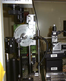
The sample can be mounted on Huber goniometer heads for manual positioning and is motorized by a goniometer allowing different movements. The position of small crystalline samples is controlled by a microscope system with CCD camera.
sketch of the sample stage (89 kB)
 picture of the goniometer from the laser direction (111 kB)
General Description
- For macromolecular crystallography the crystalline sample can rotated around a horizontal axis to take Laue images for a series of azimutal angles (phi, continuous 360 degrees).
- To allow the exact positioning of the crystal with respect to the x-ray beam (typically 200 mm square) and the laser focus, it can be mounted on goniometer heads. This allows centering the crystal within the rotation axis.
- A motorized Eulerian cradle an be used to tune the orientation in angles orthogonal to the phi circle (chi, psi, each +/- 10 degrees).
- The translation of the sample in the Y direction (horizontal, perpendicular to x-ray path) is provided by 100 mm travel with absolute encoder.
- The height of the goniometer can be changed by a Z stage, that lifts the whole setup. This is only used for the goniometer alignment at the beginning of the experiment.
Microscope
The sample positioning is recorded by a long working distance zoom microscope, that presently watches the interaction point from above and is equipped with a colour CCD camera and frame grabbing system. A future setup will allow more flexibility with the positioning of the microscope.
The new microscope can view the sample from the same direction as the x-ray beam with a variable angle vertically. It uses a fixed magnification with several fields-of-view to choose.
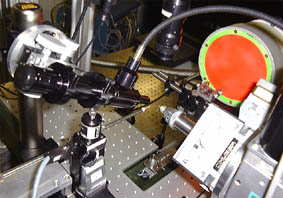
 view of the sample position (171 kB)
In the above picture one can see the x-ray beam pipe coming from left below and hitting the sample on a small goniometer head. Above the new microscope is positioned to look on the sample with an angle of 25 to 70 degrees. On the right hand side the lately installed Eulerian cradle is to be seen.


