- Home
- Users & Science
- Scientific Documentation
- ESRF Highlights
- ESRF Highlights 2013
- X-ray imaging
- Hidden anatomical features of an early tetrapod revealed by PPC-SRµCT
Hidden anatomical features of an early tetrapod revealed by PPC-SRµCT
One of the most significant events in Earth’s evolutionary history was that which produced terrestrial vertebrates (tetrapods) from water bound fish. Transformation of fins into limbs (with fingers and toes) is usually considered the most crucial step in tetrapod evolution; however, modifications of the vertebral column (and ribs) are of equal importance as the ‘backbone’ helps to both stabilise and mobilise the body during movement. The exact anatomical construction of the vertebral column in the earliest tetrapods has, however, been a point of discussion for over a century, primarily because vertebral elements remain trapped within rock matrix obscuring critical anatomical detail.
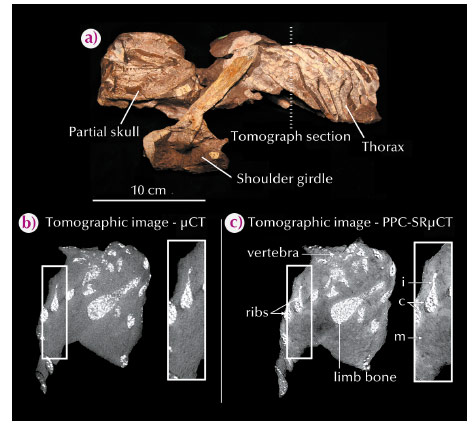
Recently, the three-dimensional skeletal anatomy of the 360 million year old stem tetrapod Ichthyostega (Figure 13a) was recreated from microcomputed tomography (µCT) scans [1]. The µCT data exposed all elements within the vertebral column and indicated the presence of new and exciting anatomical features, as well as an unexpected vertebral configuration. However, the resolution of the images was quite low, making the vertebral structures look somewhat blurry. Moreover, beam-hardening artefacts, due to the bright-white oxide inclusions within the inner spongiosa that absorbed a high proportion of the dynamic range, made the outer layer of the bone (cortex) indiscernible (Figure 13b). The resulting three-dimensional rendering of the vertebral column was of poor quality and missing detailed anatomical features necessary to make confident evolutionary inferences.
To overcome this limitation, key fossil specimens were subjected to propagation phase-contrast X-ray synchrotron microtomography (PPC-SRµCT) at beamline ID19 using a specially developed attenuation protocol for imaging large, dense fossils with metallic oxide inclusions. The attenuation protocol, detailed by Sanchez et al. [2], involves immersing fossil samples in a tube filled with aluminium or glass balls, and placing a U-shaped aluminium profiler between the beam and the sample. The net effect normalises the absorption profile by increasing the relative ratio of photons going through the densest parts of the sample, thereby providing access to a larger dynamic range of the detector. The combination of PPC-SRµCT, a polychromatic beam and the attenuation protocol, produced superior quality images as compared to traditional µCT, including substantially greater contrast and crisp anatomical details (Figure 13c).
 |
|
Fig. 13: (a) Photograph of Ichthyostega fossil material (MGUH VP 6115) showing region of the comparative tomographic slice; (b) µCT image showing a section through the thorax of Ichthyostega embedded in rock matrix, m. Notice the bright, white metallic oxide inclusions in the spongiosa, i, prevent the cortical surfaces, c, of the bones from being resolved; (c) Image at the same location as (b) obtained with PPC-SRµCT and the attenuation protocol. The image reveals improved contrast and sharp anatomical structures, with the exact cortical surfaces of the bones resolved. |
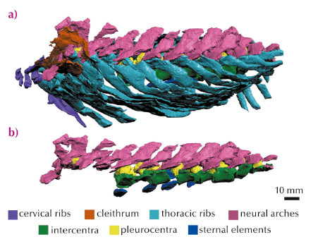
High resolution three-dimensional rendering of the PPC-SRµCT data led to the discovery of hitherto undocumented anatomical features within the vertebral column of Ichthyostega (Figure 14). These include: (1) a change in neural spine and transverse process orientation along the column, indicating specialisation of axial musculature; (2) the first evolutionary appearance of a sternum, which would have strengthened the ventral region of the ribcage perhaps permitting the animal to balance its body weight on its chest; (3) an unusual posterior-to-anterior ossification sequence, a pattern in opposition to Hox gene expression patterns and a developmental trend previously only documented in fish; (4) a complete rearrangement of the bones within each vertebral unit, a significant evolutionary result which culminated in a redefinition of the tetrapod plesiomorphic state.
 |
|
Fig. 14: Thorax region of Ichthyostega (MGUH VP 6115) rendered from PPC-SRµCT data. (a) Lateral view of whole specimens; (b) Lateral view of vertebral elements only. Rock matrix removed to highlight bony structures. Anterior towards left. Scale bar, 10 mm. |
Overall, this collaborative project added new and valuable anatomical data that has helped to redefine and elucidate the fish-tetrapod evolutionary transition. It also advanced fossil imaging techniques, opening the possibility for future groundbreaking discoveries.
Principal publication and authors
S.E. Pierce (a,b), P.E. Ahlberg (c), J.L. Molnar (b), S. Sanchez (c,d), P. Tafforeau (d), J.R. Hutchinson (b) and J.A. Clack (a), Nature 494, 226–229 (2013).
(a) University Museum of Zoology, Department of Zoology, University of Cambridge (UK)
(b) Department of Comparative Biomedical Sciences and Structure & Motion Laboratory, The Royal Veterinary College, Hatfield (UK)
(c) Subdepartment of Evolution and Development, Department of Organismal Biology, Evolutionary Biology Centre, Uppsala University (Sweden) (d) ESRF
References
[1] S.E. Pierce, J.A. Clack and J.R. Hutchinson. Nature 486, 523–526 (2012).
[2] S. Sanchez, V. Fernandez, S.E. Pierce and P. Tafforeau, Nat. Protocols 8, 1708–1717 (2013).


