- Home
- Users & Science
- Scientific Documentation
- ESRF Highlights
- ESRF Highlights 2017
- Structure of materials
- 3D reconstruction of nanowire bending and lattice tilt using X-ray diffraction
3D reconstruction of nanowire bending and lattice tilt using X-ray diffraction
Strained core-shell nanowires have been investigated with nanofocused scanning X-ray diffraction. The bending and twisting of the nanowires was reconstructed and quantified in three dimensions. In one nanowire the shell lattice was found to be tilted with respect to the core, and the azimuthal orientation of the tilt changes along the axis.
Semiconductor nanowires have shown promising performance in a wide array of applications, such as solar cells, electronics and energy storage. A particular category of these is core-shell nanowires, which are created by first growing an axial core nanowire and then changing the conditions to promote shell growth. A lattice mismatch between core and shell creates a strained heterostructure, which can enhance carrier mobility and reduce surface recombination. While the interplay between lattice mismatch, strain and defect formation is quite well understood in strained thin film heterostructures, the thin nanowires can also accommodate strain by bending.
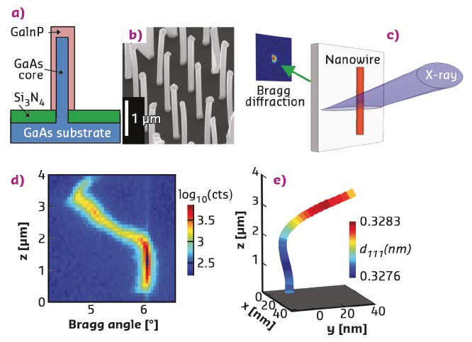
In this study, nano-XRD investigations were carried out of two strained GaAs-GaInP core-shell nanowires (Figure 126a, b). The nanowires were deposited from their growth substrate onto a Si3N4 membrane. The X-ray beam from the ID11 beamline was focused to a size of 100 nm vertically and 6 µm horizontally. Nanowires with vertical orientation, orthogonal to the beam, were chosen and aligned in the focus. Scanning nano-XRD was performed by acquiring rocking curves, that is, small scans in rotation around the [111] Bragg angle, at points with 100 nm distance along the nanowire axis (Figure 126c). The scattering of the [111] reflection was mapped into reciprocal space, and the position of the Bragg peak was evaluated as a function of the axial coordinate z.
 |
|
Fig. 126: Scanning X-ray nanodiffraction of nanowire A. a) Drawing of the GaAs-GaInP core-shell nanowire structure. b) SEM of the as-grown nanowires. c) Drawing of the experiment. d) Total counts in the Bragg peak on the detector, vs Bragg angle and z. e) 3D reconstruction of the nanowire. Note that the x and y scales are in nm and the z scale is in µm. |
Nanowire A exhibits a single diffraction peak, which shows a significant variation along the nanowire axis (Figure 126d). A perfectly straight core-shell nanowire with constant composition would show a single vertical line in such a plot, or possibly two vertical lines for a sufficiently large lattice mismatch. The Bragg angle can vary due to variations in lattice plane distance, but also due to real-space tilting of the lattice. Since the [111] planes are orthogonal to the nanowire axis, the scattering vector is locally parallel to the tangent of the single-crystal nanowire, and the contour of the nanowire can be reconstructed by stepwise integration [1]. The 3D reconstruction shows that the nanowire is strongly bent in the upper half, while the lower half is straight (Figure 126e).
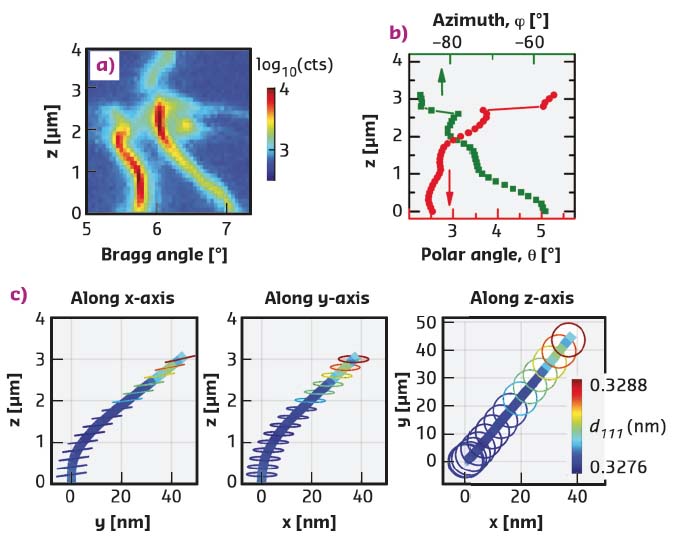
The second nanowire, B, shows two distinct Bragg peaks along most of its length (Figure 127a). The split in the Bragg angle is over one degree at the base of the nanowire, which is far more than the lattice mismatch could create. The reason is a difference in orientation of the core and shell lattices. The difference was quantified and converted to a spherical coordinate system, with one polar (Φ) and one azimuthal (θ) angle (Figure 127b). To visualise the complex crystal structure, the shape of the core was first reconstructed as nanowire A, and then circles were added to represent the orientation of the shell, with the colour showing the lattice plane distance (Figure 127c). The tilt of the planes in the plot was magnified for better visibility.
 |
|
Fig. 127: Scanning X-ray nanodiffraction of nanowire B. a) Total counts of the Bragg peak on the detector, as a function of Bragg angle and z. b) The angles between the scattering vectors of the shell and the core, vs. z. c) 3D model of the nanowire, projected along the principal axes. |
The 3D reconstruction shows that nanowire B bends but, unlike nanowire A, it exhibits no twisting. The magnitude of the bending is similar to nanowire A. The polar θ angle represents a lattice tilt between shell and core, which increases from θ = 2° at the base to θ = 5° at the top. The relative azimuthal orientation of the tilt changes from about φ = -87° near the base, to φ = –57° at the top. Thus, the shell lattice twists around the core.
These results demonstrate how scanning nano-XRD can reveal striking differences between individual nanowires, as well as within single nanowires, which would be invisible with a lower-resolution method.
Principal publication and authors
Bending and twisted lattice tilt in strained core-shell nanowires revealed by nanofocused X-ray diffraction, J. Wallentin (a, b), D. Jacobsson (b), M. Osterhoff (a), M.T. Borgström (b) and T. Salditt (a), Nano Letters 17, 4143-4150 (2017); doi: 10.1021/acs.nanolett.7b00918.
(a)University of Göttingen (Germany)
(b) Lund University (Sweden)
References
[1] J. Wallentin et al., Adv. Mater. 28 (9) (2016).


