- Home
- Users & Science
- Scientific Documentation
- ESRF Highlights
- ESRF Highlights 2017
- Structure of materials
- Seeing inside a growing catalyst
Seeing inside a growing catalyst
The formation and growth mechanism of hollow PtNi/C nanoparticles was unravelled using scanning transmission electron microscopy coupled to energy-dispersive X-ray spectroscopy, and operando high-energy wide-angle X-ray scattering. Following their formation, Ni-rich/C nanoparticles were rapidly enclosed in a Ni<sub>x</sub>ByO<sub>z</sub> shell and then slowly transformed into hollow PtNi/C nanoparticles.
Manipulating mono or bimetallic nanocrystallites at the atomic scale is pivotal to many domains of science. A textbook case concerns the unique optical, photo-thermal, paramagnetic and electrocatalytic properties of hollow metal nanoparticles. Indeed, in the presence of the oscillating electromagnetic field of a beam of light, the free electrons of many metal nanoparticles (in particular Au, Ag and Cu) oscillate coherently with respect to the positively charged metallic lattice, resulting in strong scattering and absorption. Bimetallic hollow nanoparticles have also demonstrated potential in the diagnosis of diseases, drug delivery and, more recently, in low temperature fuel cell electrocatalysis. However, the details and the kinetics of formation and growth of these peculiar nanomaterials remain unexplored.
Here, a combination of operando wide-angle X-ray scattering (WAXS) on ID31, scanning transmission electron microscopy (STEM) and X-ray energy dispersive spectroscopy (X-EDS) was used to reveal the formation and growth mechanism of hollow PtNi/C nanoparticles and provide the time scale at which the elementary steps of the synthesis occurred.
To synthesise hollow PtNi/C nanoparticles, a sodium borohydride solution (NaBH4, a very strong reductant) was added in a dropwise manner to a solution containing the Pt and Ni salt precursors and the high surface area carbon (Vulcan XC72). For STEM/X-EDS analyses, the synthesis was conducted in a glass reactor and sampling was performed at different time intervals after addition of the first drop of the NaBH4 solution. The same experimental protocol was used for operando WAXS measurements but a poly(methyl methacrylate) cuvette served as reactor.
 |
|
Fig. 115: X-ray energy-dispersive spectroscopy elemental maps of the different nanostructures forming during the synthesis of hollow PtNi/C nanoparticles. The images are recorded t = 1, 2, 3, 4, 20, 40 and 60 minutes after the addition of the first drop of the NaBH4 solution. Ni atoms and Pt atoms are represented in green and red, respectively. |
STEM/X-EDS elemental maps (Figure 115) and X-ray photoelectron spectroscopy analyses revealed that Ni/C nanoparticles around 5-10 nm in size formed first, before being embedded in a NixByOz shell (t = 2 min). This shell then served as a sacrificial template for the deposition of Pt2+ ions via a combination of chemical reduction and galvanic replacement (an electrochemical process in which a metallic phase is spontaneously oxidised by the ions of another metal having a higher reduction potential). Finally, in the time period 20 ≤ t ≤ 60 min, the nanoscale Kirkendall (a spontaneous vacancy-mediated interdiffusion mechanism occurring in binary alloys or core@shell materials) contributed to the transformation of the solid Ni-rich core@Pt-rich shell nanoparticles into hollow alloy PtNi/C nanoparticles.
 |
|
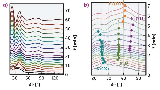
Fig. 116: Operando wide angle X-ray scattering patterns recorded during the synthesis of hollow PtNi/C nanoparticles. The time (t) corresponds to the time elapsed since the addition of the first drop of the NaBH4 solution. Representative patterns are presented in (a), whereas (b) highlights the position and the assignment of the peaks in the 0 ≤ t ≤ 6.5 min time period. Note that the raw data (energy 60.0 keV corresponding to λ = 0.207 Å) were previously converted into Cu Kα radiation (λ = 1.5406 Å) for the sake of comparison with classical X-ray diffraction. |
This mechanism of formation was confirmed using operando WAXS. Diffraction from the [002] planes of the carbon support, monoclinic NiB3 and/or tetragonal Ni2B, Ni(III) and Pt(III) appeared sequentially (Figure 116). The disappearance of the Ni(III) signal at t ≥ 6 min confirmed that the Ni cores were corroded, or alloyed with Pt. Later in the reaction, the reflections ascribed to Pt increased in intensity, indicating enrichment in Pt in agreement with the STEM/X-EDS elemental maps displayed in Figure 115.
Beyond their interest for catalysis and electrocatalysis, the results provide for the first time a powerful methodology, capable of identifying the formation and growth mechanism of preferentially-shaped metal nanoparticles, core@shell nanoparticles, onion-like nanoparticles, Janus nanoparticles or a combination of all of these.
Principal publication and authors
Atomic scale snapshots of the growth mechanism of hollow PtNi/C nanocatalysts for oxygen reduction reaction, R. Chattot (a,b), T. Asset (a,b), J. Drnec (c), P. Bordet (d,e), J. Nelayah (f), L. Dubau (a,b) and F. Maillard (a,b), Nano Lett. 17, 2447-2453 (2017); doi: 10.1021/acs.nanolett.7b00119.
(a) Université Grenoble Alpes, LEPMI, Grenoble (France)
(b) CNRS, LEPMI, Grenoble (France)
(c) ESRF
(d) Université Grenoble Alpes, Institut Néel, Grenoble (France)
(e) CNRS, Institut Néel, Grenoble (France)
(f) Université Paris Diderot, Sorbonne Paris Cité, CNRS, Laboratoire Matériaux et Phénomènes Quantiques, UMR 7162, F-75013, Paris (France)


