- Home
- Users & Science
- Scientific Documentation
- ESRF Highlights
- ESRF Highlights 2017
- Structure of materials
- X-ray ghost imaging using individual synchrotron pulses
X-ray ghost imaging using individual synchrotron pulses
Ghost imaging is an indirect method in which most of the X-rays used for the experiment never actually interact with the sample. The method uses two copies of the same speckled beam and the image is retrieved indirectly by correlating the two measured signals. This experiment could lead to the development of low-dose medical X-ray diagnostics and diffraction imaging at free electron lasers.
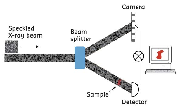
The time structure of the ESRF synchrotron beam has been used to demonstrate, for the first time ever, the X-ray version of ghost imaging, carried out at beamline ID19. Ghost imaging is an indirect imaging method that has received considerable attention in recent years [1]. In its simplest form, ghost imaging requires two identical copies of a structured (speckled) beam, as illustrated in Figure 122. One copy is directed on the sample and only the total intensity (not the image) transmitted or scattered by the specimen is recorded by a bucket (point) detector. The second, empty, beam is imaged by a pixel array detector, to form the reference image. In this way the sample image is never recorded directly. Remarkably though, the sample image can be reconstructed by repeating the experiment many times, each time with a different speckle pattern. The image is retrieved by correlating the bucket signals and the reference images. This can be done with an electronic correlator or, as in the case of this experiment, working offline on the whole data sequence.
 |
|
Fig. 122: Schematic diagram of a ghost-imaging experiment. |
Combining the special ‘4-bunch’ filling mode of the ESRF with the intense X-ray pulses produced by the undulators at beamline ID19, the first proof-of-principle demonstration of ghost imaging in the hard X-ray spectral range was achieved. In 4-bunch mode, the temporal separation between the bunches is approximately 704 ns, corresponding to a frequency of about 1.42 MHz. In this experiment, the X-ray pulses were monochromatised to an energy of 20 keV by a pair of silicon crystals. The beam was then split using a thin silicon crystal in Laue diffraction, to produce the two identical copies required by the ghost imaging protocol. The sample (an X-ray opaque Cu wire) was inserted into one of the beams and both beams were detected using the same camera frame.
An ultra-fast camera was used to image the individual X-ray pulses, for an average of only a few consecutive pulses. In this way the characteristic shot noise of the synchrotron emission process is dominant and the beam contains natural speckles. In this proof-of-concept experiment, the bucket signal was calculated by summing the intensity measured in the sample beam and correlated with the reference beam image instead of using a separate bucket and reference detector.
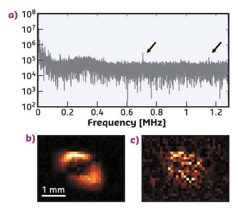
The power spectrum of the bucket signal is shown in Figure 123a. The sharp peak at 0.71 MHz corresponds to half the storage ring frequency. Low frequency components of the spectrum arise from mechanical vibrations of the optics (monochromator and crystal beam splitter), which are problematic. Vibrations cause small changes in the crystal orientation, which produce intensity variations in both the diffracted and transmitted beam. Such variations are actually anti-correlated and therefore obscure the genuine speckle correlation coming from the electron bunch structure.
To eliminate the effect of mechanical vibrations, Fourier filtering of the spectrum of both reference and bucket signals was performed. The true speckle correlation was isolated by windowing the 0.71 MHz peak (equivalent to taking the average of two pulses) in both reference and bucket. Figure 123b shows the recovered ghost image of the wire, using the filtered spectrum. Figure 123c shows the ghost image obtained when the window selects a nearby spectral region instead of the 0.71 MHz peak. In this latter case the speckle correlation is not present and the sample image is not recovered.
 |
|
Fig. 123: a) Power spectrum of the bucket signals. Two peaks are visible in the high frequency part (marked by arrows). The actual storage ring frequency (1.42 MHz) was not resolved due to the insufficient temporal resolution of the camera. b) Ghost image of a copper wire measured by windowing the 0.71 MHz frequency component. c) By windowing the ‘wrong’ frequency component (in this case 0.8 MHz) the ghost image is not retrieved. |
The experiment confirms that shot noise produced in the synchrotron emission of isolated electron bunches can be successfully used for correlation imaging experiments. This result might inspire new ideas on single molecule diffraction imaging at FELs. By reducing the dose of the bucket beam, a “diffraction without destruction” protocol using ghost imaging [2] could be devised.
X-ray ghost imaging has other potential applications that include spectroscopy, such as X-ray fluorescence, where an imaging detector generally cannot be used. Also, it could be highly beneficial for low dose radiology as most of the photons in a ghost imaging experiment never interact with the sample.
Principal publication and authors
Experimental X-ray ghost imaging, D. Pelliccia (a, b), A. Rack (c), M. Scheel (d), V. Cantelli (e, c) and D. Paganin (f), Phys. Rev. Lett. 117, 113902 (2016); doi: 10.1103/PhysRevLett.117.113902.
(a) Instruments & Data Tools Pty Ltd (Australia)
(b) RMIT University, Melbourne (Australia)
(c) ESRF
(d) Synchrotron Soleil, Gif-sur-Yvette (France)
(e) Helmholtz-Zentrum Dresden-Rossendorf, Dresden (Germany)
(f) Monash University, Melbourne (Australia)
References
[1] B.I. Erkmen and J.H. Shapiro, Adv. Opt. Phot. 2, 405-450 (2010).
[2] Z. Li et al., arXiv:1511.05068 (2015).


