- Home
- News
- Spotlight on Science
- X-ray spectroscopy...
X-ray spectroscopy reveals iron spin state in oxygenated hemoproteins
13-11-2017
Proteins carrying a heme cofactor are essential for biological oxygen management. Advanced X-ray spectroscopy experiments have now established an effective intermediate-spin iron centre in myoglobin and hemoglobin. This has clarified the long-debated Fe-O2 bonding situation in reversible oxygen transport.
Share
Aerobic life on earth requires efficient management of molecular oxygen (O2). Nature has developed proteins that bind an iron-porphyrin (heme) cofactor as versatile O2 managers. Such hemoproteins, in particular in animals and humans, have been extensively studied, for example, establishing cooperative versus non-cooperative O2 binding to heme iron in tetrameric hemoglobin and monomeric myoglobin, respectively. Today, rising interest in this topic stems from the ongoing discovery of globins with broad functionality in plants and microorganisms.
Three physiologically-relevant heme species can be distinguished in myoglobin and hemoglobin: the ligand-free (deoxy), carbon-monoxide inhibited (carboxy), and O2-bound (oxy) forms. Paramagnetic behaviour has established high-spin Fe(II) in deoxy whereas carboxy is diamagnetic due to low-spin Fe(II). For oxy, a diamagnetic cofactor has been proven. A controversy has remained on the nature of the iron-oxygen bonding in myoglobin and hemoglobin, dating back to the pioneering work of Pauling, Monod, and Perutz that began in the 1930's [1]. Pauling suggested a neutral singlet O2 bound to a formal low-spin Fe(II), McClure and Goddard suggested a triplet O2 and an intermediate-spin Fe(II) in an ozone-like configuration, and Weiss suggested an Fe(III) coupled to a superoxo (O2-) ligand [2].
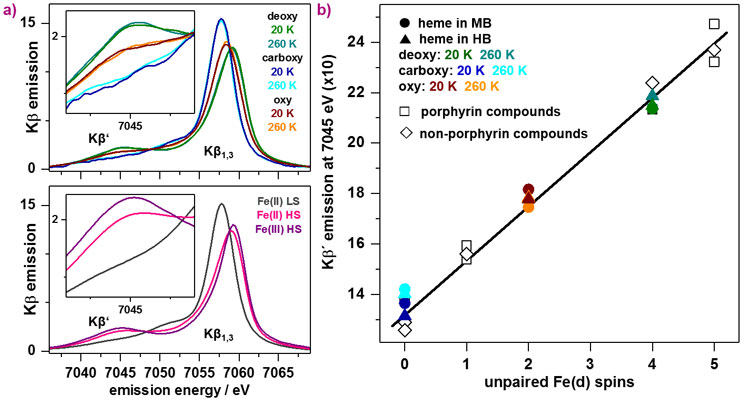
Core level spectroscopy is a powerful method for the electronic structure analysis of transition metal complexes. X-ray emission spectroscopy of the Kβ lines probes the occupied levels via electronic decay from metal 3p to 1s levels (main line). 3p-3d spin-coupling splits the Kβ main-line emission into the Kβ’ and Kβ1,3 features and the Kβ’ intensity is a measure of metal spin state and bonding covalency. X-ray spectroscopy on hemoproteins in solution and at physiological temperature is challenging due to radiation-induced modifications and low metal concentration.
At beamline ID26, myoglobin and hemoglobin proteins were studied in their deoxy, carboxy and oxy forms, as well as synthetic porphyrin compounds with low-spin or high-spin Fe(II) or Fe(III) centres. Kβ main-line emission spectra were collected at cryogenic (20 K) or near-ambient (260 K) temperatures (Figure 1). Significant line shape differences between the protein-bound heme species reflect iron spin and redox state differences. The temperature-invariant behaviour of the spectra proved the absence of spin crossover and thereby the physiological relevance of the determined spin states. Plotting of the Kβ’ intensities versus the formal number of unpaired Fe(d) spins revealed a linear relation. The mean Kβ’ amplitude for oxy heme was in best agreement with an Fe(d) spin count of two. These results strongly suggest that the high-spin Fe(II) in deoxy myoglobin and hemoglobin is converted to an effective intermediate-spin iron species upon oxygen binding to the metal.
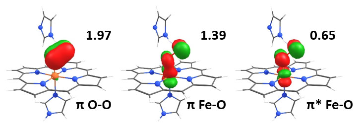
Quantum chemical ab-initio calculations confirmed the experimentally determined number of unpaired Fe(d) electrons. For the high-spin Fe(III) in deoxy heme, five single-occupied molecular orbitals with Fe(d) character were found and the low-spin Fe(II) in carboxy heme showed no unpaired electrons. For oxy heme, the unpaired electron density of two is the result of partial electron occupation of two orbitals representing bonding (π) and antibonding (π*) Fe-O interactions. Additional correlation with the π orbital of O2 yields an electronic configuration in oxy heme similar to the one in ozone (Figure 2).
 |
|
Figure 2. Iron-oxygen bonding in heme. Comparison of molecular orbitals with non-integer electron occupation (numbers) from quantum chemical (complete active space self-consistent field) calculations shows that the effective intermediate-spin behaviour of the ferrous iron in oxy heme originates from a delocalised iron-oxygen bonding situation similar to that of ozone. |
This work has helped to resolve the longstanding controversy concerning the iron-oxygen interaction and metal spin state in oxygen transporting proteins. It was achieved using quantitative correlation of advanced X-ray absorption and emission spectroscopy experiments and quantum chemical calculations. The situation in oxygenated hemoglobin and myoglobin merges the classical models into a unifying view of the Fe-O2 bonding, which features limited radical character and delocalised backbonding stabilisation of the ligand as prerequisites in reversible biological O2 transport. A framework has been provided to understand the tuning of hemoproteins for their numerous functions ranging from O2 sensing and transport over activation chemistry to catalysis.
Principal publication and authors
Effective intermediate-spin iron in O2-transporting heme proteins, N. Schuth (a), S. Mebs (a), D. Huwald (b), P. Wrzolek (c), M. Schwalbe (c), A. Hemschemeier (b), M. Haumann (a), Proc. Natl. Acad. Sci. U. S. A. 114, 8556-8561 (2017); doi: 10.1073/pnas.1706527114.
(a) Freie Universität Berlin, Department of Physics, Berlin (Germany)
(b) Ruhr-Universität Bochum, Department of Plant Biochemistry, Bochum (Germany)
(c) Humboldt-Universität zu Berlin, Department of Chemistry, Berlin (Germany)
References
[1] K.P. Kepp, Coord. Chem. Rev. 344, 363-374 (2016).
[2] K.L. Bren, R. Eisenberg and H.B. Gray, Proc. Natl. Acad. Sci. U. S. A. 112, 13123-13127 (2015).


