- Home
- News
- General News
- Powders show their...
Powders show their strength
09-10-2007
Growing a single crystal of a protein can be very difficult. Thanks to recent developments, a powder sample may be enough to solve a structure. Researchers from the European Molecular Biology Laboratory (EMBL-Hamburg) and the ESRF have successfully used the powder diffraction technique to determine the structure of the second SH3 domain of ponsin, which plays an important role in muscle differentiation. The results are comparable to those obtained by single crystal techniques and demonstrate the power and the potential applicability of the powder technique in structural biology.
Share
The knowledge of the 3D structure of proteins and protein assemblies provides valuable information in health science research and biotechnological applications. X-ray crystallography is the method researchers use to visualise the structures of macromolecules. However, it can be challenging, even impossible, to grow crystals that are suitable for standard single crystal measurements. Often, macromolecules such as amyloid fibrils, supramolecular assemblies or even proteins are particularly hard to crystallise. On the other hand, the preparation of a powder sample – comprising a large number of very tiny randomly oriented crystallites– is much less demanding. Powders can be prepared for many samples simply by batch precipitation. Nevertheless, analysing data from powders is more complicated, and generally limited to comparably simple crystal structures.
The two teams from the ESRF and the EMBL, focused on the study of an SH3 domain of ponsin, a protein involved in muscle differentiation. Furthermore, the SH3 motif is one of the most abundant found in a large variety of signalling processes thus linked to several diseases and is therefore an important therapeutic target for drug design.
 |
|
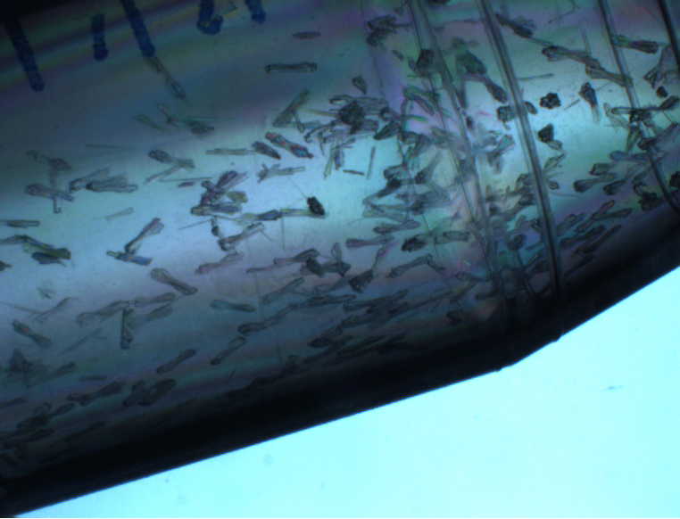
The sample. After purification, the SH3.2 domain spontaneously formed a microcrystalline material suitable only for powder diffraction measurements. |
|
|
|
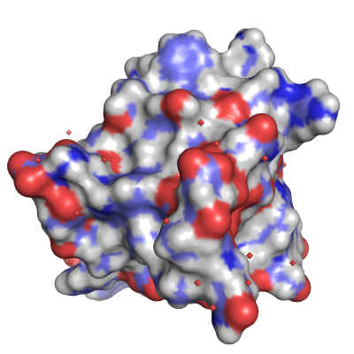
The structure. Electrostatic potential representation of the 67 amino acid SH3 domain revealed from powder diffraction. The identified water molecules are shown as red spheres. |
Upon sample purification the specific SH3-domain protein sample spontaneously formed a suspension of microcrystals (see image “the sample”). This material was suitable only for powder diffraction and therefore it was brought to the European Synchrotron Radiation Facility in Grenoble where scientists collected very high quality data. The molecular arrangement of the domain was gradually unravelled from the powder diffraction patterns using the joint expertise of the two research teams.
They created novel data processing strategies drawing on both traditional single crystal and powder methods. For example, radiation damage or modest changes in sample preparation conditions lead to small shifts of the diffraction peak positions in a powder pattern. Overlapped diffraction peaks can then be better resolved by combining multiple patterns and exploiting these differences. The final result was a powder dataset which approaches single crystal quality (dmax ~ 2.4 Å).
In order to start the analysis, scientists used a similar SH3 domain as a structural model. They determined the orientation and position of the protein into the unit cell via molecular replacement. The structure was refined initially to produce electron density maps which were then employed to trace the main amino acid chain alterations and find the missing atoms. The structure was completed via a multiple-data-set Rietveld fit.
To the team’s surprise, they could even detect water molecules in electron density maps (see image “the structure”); a substantial observation from powder diffraction data. These results were later verified by single crystal measurements, as the conditions for growing a single crystal of the protein, suitable for structural analysis, were subsequently discovered.
The study on this SH3 domain represents a step forward in the development of modern X-ray diffraction methods for micro- and nano-crystalline samples. “We believe that powder methods may have a significant impact in the fields of structural biology and biomaterial sciences – just as it has had in solid-state and structural chemistry over the last twenty years”, explains Irene Margiolaki, first author of the paper. “The structure of this important protein confirms that powder diffraction is now a technique ready to tackle genuine biological problems”, she concludes.
Reference:
I. Margiolaki, J. P. Wright, M. Wilmanns, A. N. Fitch & N. Pinotsis, "Second SH3 Domain of Ponsin Solved from Powder Diffraction", J. Am. Chem. Soc., 129 (38), 11865 -11871, 2007.


