- Home
- News
- General News
- Unravelling Elastin,...
Unravelling Elastin, nature’s amazing spring
10-03-2011
Scientists have used three synchrotrons, including the ESRF, to reveal the shape of the protein that gives human tissues their elastic properties. This discovery might lead to the development of new synthetic elastic polymers.
Share
An international team of researchers from the UK, Australia, USA and Europe have solved the complex structure of tropoelastin, the main component of elastin. The team used experimental stations at Diamond Light Source in Didcot (UK), the European Synchrotron Radiation Facility (ESRF) in Grenoble, France, and the Advanced Photon Source (APS) in Chicago, USA, to make this breakthrough, the result of more than a decade of international collaboration. Their findings are published in the March issue of science journal PNAS.
Elastin allows tissues in humans and other mammals to stretch, for example when the lungs expand and contract for respiration or when arteries widen and narrow over the course of a billion heart beats.
“All mammals rely on elastin to provide their tissues with the ability to stretch and then return to their original shape,” said researcher Dr Clair Baldock, from the University of Manchester’s Wellcome Trust Centre for Cell Matrix Research. “This high level of physical performance demanded of elastin vastly exceeds and indeed outlasts all human-made elastics. It is the co-ordinated assembly of many tropoelastins into elastin that gives tissues their stretchy properties and this exquisite assembly helps to generate elastic tissues as diverse as artery, lung and skin.
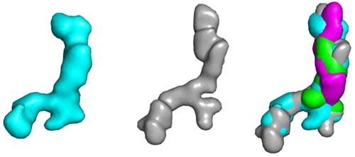
“We discovered that tropoelastin is a curved, spring-like molecule with a ‘foot’ region to facilitate attachment to cells. Stretching and relaxing experiments showed that the molecule had the extraordinary capacity to extend to eight-times its initial length and can then return to its original shape with no loss of energy, making it a near-perfect spring.”
Professor Nick Terrill, Principal Beamline Scientist for the Non-crystalline diffraction beamline (I22), the experimental station used at Diamond said: “The synchrotron technique used was biological solution scattering and we’re currently working on improving I22’s capabilities in this area. We are collaborating with the ESRF and the European Molecular Biology Laboratory (EMBL) Hamburg to develop automation and automated data analysis techniques for solution scattering in biology so we’re hoping to do a lot more of this kind of research on I22 in the future.”
“The full length protein is very flexible and it is not likely to be amenable to crystallographic studies. Small-angle-X-ray-scattering measurements carried out at ESRF beamline ID02 provided crucial initial data,” adds Theyencheri Narayanan, Head of the Structure of Soft Matter Group at the ESRF.
 |
|
Structure of the full-length tropoelastin calculated from X-ray scattering (left), neutron scattering (middle) and a representation of the two combined (right). |
Elastics are used in applications as diverse as clothing, vehicles, tissue engineering and even space travel, so understanding how the structure of tropoelastin creates its exceptional elastic properties could enable the development of synthetic ‘elastin-like’ polymers with potentially wide-ranging applications and benefits.
Initiator and research project leader Tony Weiss, Professor in the School of MolecularBioscience, The University of Sydney, added: “Tropoelastin’s extraordinary capacity to extend to eight-times its initial length and then return to its original shape, with no loss of energy, is nature showing us how to make an ideal nanospring.”
Reference: Clair Baldock et al., Shape of tropoelastin, the highly-extensible protein that controls human tissue elasticity, PNAS March 2011 published online 28 February 2011, DOI: 10.1073/pnas.1014280108
Top image: Structure of tropoelastin.


