- Home
- News
- Spotlight on Science
- X-ray speckle tracking:...
X-ray speckle tracking: a new phase-sensitive imaging technique
14-06-2012
A new phase-sensitive technique has been developed at the ESRF to quantitatively map the phase shift induced by a low absorbing sample within a coherent X-ray beam. It permits imaging the interior of a sample with micrometre scale or better resolution and offers many advantages over earlier methods such as minimalist setup, high sensitivity and the possibility of investigating dynamic samples.
Share
Soft matter, low density materials, can be imaged with higher sensitivity than ever before using phase sensitive techniques thanks to X-ray beams with a more spherical wavefront and better coherence gained recently through improvements to the X-ray source of the ESRF.
A team of scientists working at beamline BM05 has developed a new phase sensitive technique called X-ray Speckle Tracking (XST) that offers many advantages over existing propagation or grating based techniques. It has a very simple setup that involves a membrane of small grains whose dimensions are only statistically known, for example a biological filtering membrane or abrasive paper (Figure 1), which creates a random pattern on a camera when illuminated with X-rays. This pattern is generated because the features in the membrane create local modifications in the incoming beam wave front: the beam is split into various microwavelets creating a random interference pattern, a granular structure known in optics as ‘speckle pattern’.
 |
Figure 1. Examples of membrane and abrasive paper used in the phase imaging setup. |
The new XST technique results from the combination of two concepts. The first concept is based on the different propagation behaviour for speckles generated by X-rays and by visible light. With visible light the speckle pattern changes quickly upon space propagation, while for X-rays the speckle pattern remains the same over a distance that can vary from a few millimetres to several metres, depending on the photon energy and the beam divergence [1]. The second concept is the ability to track speckle grain subsets between images taken either at two different distances or at two different moments in time. Since this measuring principle is similar to the field of mechanical strain measurements in material science [2], specific algorithms used there were adapted to the XST case. From the displacement of the speckle features between images, it is possible to infer the propagation direction of the X-rays carrying each of the speckle grains, and hence the X-ray paths. Knowing the ray trajectories, which is given by the wave front and the phase of the beam, one can then draw up a map of the refractive index of the sample under investigation.
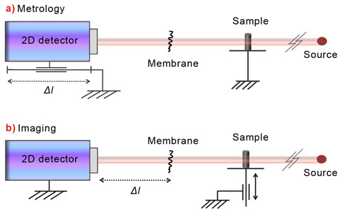
The simplicity of the technique is one of its most appealing features. Compared to other techniques, it has reduced requirements on the degree of coherence, a sensitivity and a spatial resolution higher than the instruments commonly used such as grating interferometers and Hartman sensors. Moreover, when using the imaging configuration, it provides two dimensional quantitative information about the phase shift induced by the sample for a single exposure to X-rays (Figure 2).
 |
|
Figure 2. Experimental configurations for: a) Online metrology; b) Imaging. |
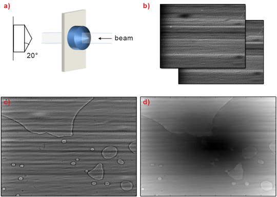
The method can be used for the phase imaging of small and low density samples thanks to its high sensitivity (Figure 3). In addition, as only one of the two images of the process has to be taken with the sample inserted into the beam, the technique is suitable for the imaging of dynamic samples.
Another field of application is online metrology, for instance for the characterisation of synchrotron beams and optics. An interesting perspective would also be to employ the technique in the new XFEL instruments emerging worldwide, because of the high spatial resolution this method provides and its unique ability to exploit each X-ray beam shot generated by such a pulsed source.
Acknowledgement
The authors thank I. Zanette, T. Weitkamp, P. Coan and P. Diemoz for providing the samples used in their experiments.
Principal publication and authors
Two-dimensional X-ray beam phase sensing, S. Berujon (a,b), E. Ziegler (a), R. Cerbino (c), L. Peverini (a,d), Physical Review Letters 108, 158102 (2012).
(a) ESRF
(b) Diamond Light Source Ltd (UK)
(c) Università degli Studi di Milano (Italy)
(d) Present address: SESO, Aix-en-Provence (France)
References
[1] R. Cerbino, L. Peverini, et al., Nature Physics 4, 238-243 (2008).
[2] B. Pan, K. Qian, et al., Measurement Science and Technology 20, 062001 (2009).
Top image: Speckle-modulated X-ray wavefront.


