- Home
- News
- Spotlight on Science
- Bone ultrastructure...
Bone ultrastructure resolved in 3D by phase nanotomography
09-10-2012
Bone tissue was imaged at the subcellular level with synchrotron X-ray phase nanotomography. Images at the ultrastructural level have previously only been available with electron imaging techniques, such as scanning or transmission electron microscopy, thus in 2D. In this study, the bone ultrastructure was imaged in 3D with a 60 nm voxel size. Several ultrastructural properties could be visualised in 3D for the first time: the collagen fibre orientation, the mineralisation on the nanoscale, and the morphology of the imprint of the cellular network. Since bone strength and failure are increasingly thought to be due to ultrastructural properties, this technique makes possible new studies to understand and cure bone diseases.
Share
Bone is a hierarchically organised material with remarkable capabilities to maintain and repair itself. However, due to aging or disease, bone becomes fragile, leading to fracture. This is a severe public health concern with the aging population. Understanding the mechanisms that determine bone strength and fragility is the prerequisite for advances in bone tissue engineering and disease treatment. For example, the role of the osteocyte, a bone cell embedded in the bone matrix, in sensing damage and converting forces to biological signals has been highlighted. Despite recent advances, a clear picture of the 3D nano-organisation of bone, such as the collagen fibre orientation, has not yet been established due to lack of suitable 3D imaging techniques [1-4].
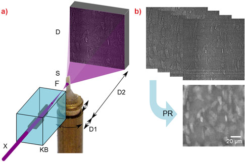
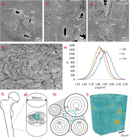
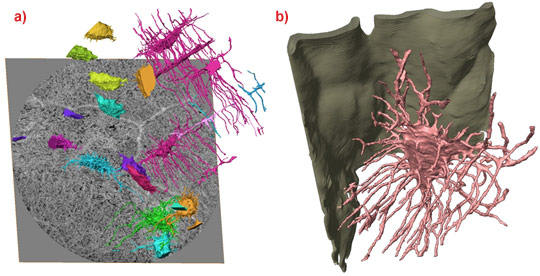
Synchrotron nanotomography provides a unique tool for the investigation of bone ultrastructure. Magnified X-ray phase nanotomography was performed at the nano-imaging station ID22NI of the ESRF (Figure 1). Researchers from INSA Lyon and the ESRF obtained 3D maps of the mass density within human cortical bone samples at 60 nm spatial resolution (Figure 2). The images reveal the organisation of the lacunae, which contain the osteocytes in living bone, and the canaliculi through which they connect. This 3D lacuno-canalicular system could be depicted over a large area including part of an osteon (the basic functional unit of bone), as well as some interstitial tissue (old bone) and the cement line separating the two types of tissue. At the cement line interface, canaliculi either turn along it and stay in the osteon, or they attach to the cement line, without traversing it. The cement line itself could also be studied in 3D for the first time (Figure 3). Previously, there was no consensus on the composition of the cement line, as some studies have shown that it contains more mineral than the surrounding tissue, while other suggested that it contains less. In this study we could directly measure the relative electron density, and we show that the cement line is hypermineralised. For the first time, the collagen organisation in the compact bone matrix could be directly observed in 3D. Here there was no clear consensus either, and several models have previously been suggested to explain the appearance of the bone matrix in 2D. The imaged samples revealed that the collagen fibres are organised in a complex manner, as a twisted plywood structure.
Magnified X-ray phase nanotomography possesses a number of unique advantages. The information content of the 3D images is exceptionally high due to the high spatial resolution and large field of view. The technique requires very little sample preparation, thus avoiding possible unintentional altering of structures. The sensitivity is exceptional and seems to outperform attenuation based full-field X-ray microscopy and ptychography [3]. As opposed to scanning techniques, it is possible to keep acquisition times short (less than two hours per sample) thus facilitating the imaging of series of samples. We believe this method will have a significant impact in osteology since it gives access to information unavailable with other techniques, while being fast and reproducible. Therefore, this work paves the way for new studies towards a better understanding of bone function, remodelling, and ultimately to new strategies in bone repair and the management of bone related diseases.
Further information
Principal publication and authors
X-Ray Phase Nanotomography Resolves the 3D Human Bone Ultrastructure, M. Langer (a,b), A. Pacureanu (a,b,c), H. Suhonen (b), Q. Grimal (d), P. Cloetens (b), F. Peyrin (a,b,e), PLoS ONE, 7: e35691 (2012).
(a) CREATIS, CNRS 5220, INSERM U1044, INSA Lyon, Université de Lyon (France)
(b) ESRF
(c) Current address: Centre for Image Analysis & SciLifeLab, Uppsala University (Sweden)
(d) LIP, CNRS, Université Paris (France)
(e) Labex PRIMES, Université de Lyon (France)
References
[1] P. Schneider et al., Bone 47, 848-858 (2010).
[2] L.F. Bonewald, J Bone Miner. Res. 26, 229-238, (2011).
[3] M. Dierolf et al., Nature 467, 436-439 (2010).
[4] A. Pacureanu, M. Langer, E. Boller, P. Tafforeau, F. Peyrin, Med. Phys. 39, 2229-2238 (2012).
Top image: Osteocyte lacunae and canaliculi rendered in 3D.


