- Home
- News
- Spotlight on Science
- X-ray Microtomography...
X-ray Microtomography of Deployment of a Cardiovascular Stent
22-12-2005
Share
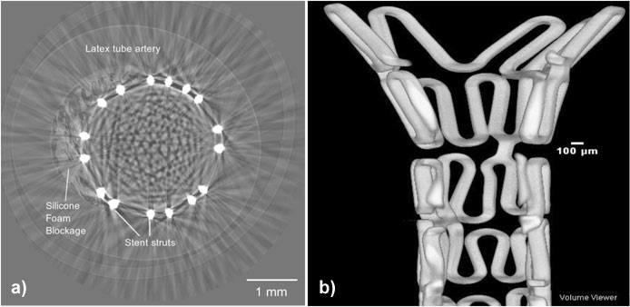
The favoured treatment for blocked coronary arteries is angioplasty, a procedure in which the blockage is cleared by balloon dilation, followed by deployment of a metallic scaffold known as a stent. The stent remains permanently in place, providing support to keep the artery open. Sometimes the success of this procedure is compromised by re-blockage of the artery by new tissue growth, due to an over-active inflammatory response triggered by balloon and stent interaction with the artery wall during surgery. Despite the introduction of drug coated stents to reduce re-stenosis, it is recognised that stent design can be improved to reduce vessel injury. However, the interaction of a stent with an artery is difficult to measure in laboratory tests because of the small device size (typically 15-20 mm long; expanded diameter 3.0 mm; strut width 0.1 mm). The objective of this experiment performed at the ESRF, beamline BM05 was to see if high resolution X-ray microtomography could be used to obtain 3D images of stent deployment within an artificial artery. The artificial artery used natural latex and silicone foam to model the artery wall and coronary plaque blockage. The stents used were R-Stents. Microtomography scans of stent deployment at various pressures were successfully obtained, with a voxel resolution of 5.3 µm. Tomography scan slices are currently being used to make various measurements of stent-artery interaction (Figure 1a). 3D reconstructions from the scans have also been generated (Figure 1b) and will be used to evaluate finite element simulations of stent deployment.
Authors
Thomas Connolley (a), Jean-Yves Buffière (b), Faisal Sharif (a), Derek Nash (a)
(a) National Centre for Biomedical Engineering Science, NUI, Galway (Ireland)
(b) Institut National des Sciences Appliquées, Lyon (France)


