- Home
- News
- Spotlight on Science
- Core-shell nanowires...
Core-shell nanowires reveal their inner structure
07-08-2009
Nature creates the most complex structures by assembling single atoms or molecules into functional building blocks. Semiconductor physics has been exploring this “self-assembly” phenomenon for several decades. An interesting example is self-assembled core-shell nanowires, where the nanowires are surrounded by a radial shell consisting of a higher band gap material. Using advanced X-ray structural characterisation techniques, scientists deduced the average shape, chemical composition, and strain field of core-shell nanowire ensembles.
Share
The wide-spread interest in semiconductor nanowires (NWs) is driven by their potential to constitute suitable building blocks for future nanoelectronic and nanophotonic devices [1]. Such applications require complex nanowire structures with either axial or radial nanowire heterostructures to provide carrier confinement and/or the waveguiding of light. Recently, increased attention has been paid to the InAs/InP core-shell system [2] from which high electron mobility nanowire field effect transistors (NWFET) have been realised [3]. The strain due to the different lattice constants of core and shell material in such radial heterostructures plays an important role [4]. This strain also induces constraints on the thickness and chemical composition of the shell for a given core, if one aims at the growth of defect-free structures required for most applications. We have investigated a series of InAs core/InAs1-xPx shell nanowires, grown by metal-organic vapour phase epitaxy on Si (111) substrates without the use of an Au catalyst [5]. X-ray diffraction reciprocal space mapping (beamline ID10B) in two X-ray scattering geometries was used for the quantitative determination of the strain field inside the nanowires. We used both coplanar high-angle diffraction (XRD), which is sensitive to the lattice parameter in the growth direction, and grazing-incidence diffraction (GID), which is very sensitive to the in-plane lattice parameters, i.e. perpendicular to the growth direction. The XRD measurements let us envision two different situations for the strain state of the investigated samples. The first is pseudomorphic growth, where the shell material matches the crystal lattice of the core at the core/shell interface. Therefore the lattice parameter in growth direction is identical for core and shell, and only one sharp peak is seen in XRD. The second is plastically relaxed growth, where defects are introduced and the shell has a different lattice parameter to the core. Several measurements show peak broadening along the growth direction, due to a distribution of lattice parameters between the shell and core lattice parameters, indicating the beginning of plastic relaxation.
To verify these conclusions and to derive quantitative results, GID measurements were taken around the (100), (200), (300) hexagonal in-plane reflections [6]. These measurements were compared with simulations to obtain information on shape, strain, and chemical composition. Beginning with finite element simulations (FEM) of a core-shell nanowire where the correct nanowire shape, the crystal orientations and the anisotropic elastic properties of the involved compounds were taken into account, we derived the displacement field inside the nanowires. From this displacement, the scattered intensity distribution in reciprocal space was calculated.
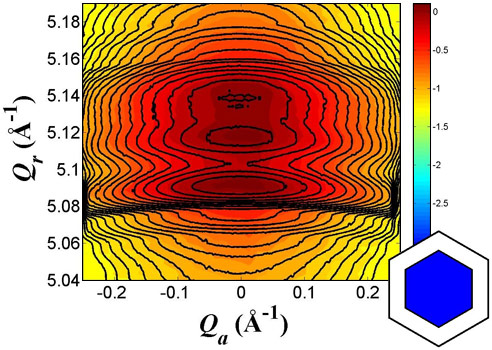
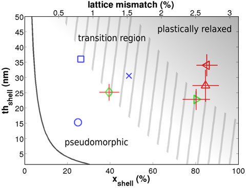
Samples with low P content and thin shells give excellent agreement between measurement and simulation, if we assume fully pseudomorphic shell growth (Figure 1). On the other hand, samples with high P content show a poor correspondence between experiment and simulation assuming pseudomorphic growth. Therefore, plastic relaxation has to be considered. Since this was not directly possible in our FEM model, we mimicked plastic relaxation phenomenologically by "cutting open" the shell to some extent to allow for additional relaxation. We got reasonable agreement between simulation and measurement using this model, however parameters like where and how deep we cut the shell could be chosen over a wide range of values. Different simulations gave slightly different parameter sets for shell thicknesses and compositions. In the resulting phase diagram of the transition from pseudomorphic to plastically relaxed growth (Figure 2) this variance is represented by the error bars.
In conclusion, from the X-ray diffraction data, we derived the structure of the core-shell nanowires concerning their strain distribution, chemical composition, shell thickness, and onset of plastic relaxation. Detailed GID experiments combined with finite element simulations lead to quantitative results, and a phase diagram of the transition from pseudomorphic to plastically relaxed growth, depending on the nanowire shell thickness and P content.
References
[1] C. Thelander, P. Agarwal, S. Brongersma, J. Eymery, L.F. Feiner, A. Forchel, M. Scheffler, W. Riess, B.J. Ohlsson, U. Gosele, L. Samuelson, Materials Today 9, 28 (2006).
[2] X. Jiang, Q. Xiong, S. Nam, F. Qian, Y. Li, C.M. Lieber, Nano Letters 7, 3214 (2007).
[3] T. Bryllert, L.E. Wernersson, L.E. Fröberg, L. Samuelson, IEEE Electron Device Letters 27, 323 (2006).
[4] N. Sköld, L.S. Karlsson, M.W. Larsson, M.E. Pistol, W. Seifert, J. Trägårdh, L. Samuelson, Nano Letters 5, 1943 (2005).
[5] B. Mandl, J. Stangl, T. Mårtensson, A. Mikkelsen, J. Eriksson, L.S. Karlsson, G. Bauer, L. Samuelson, W. Seifert, Nano Letters 6, 1817 (2006).
[6] T. Mårtensson, J.B. Wagner, E. Hilner, A. Mikkelsen, C. Thelander, J. Stangl, B.J. Ohlsson, A. Gustafsson, E. Lundgren, L. Samuelson, W. Seifert, Advanced Materials 19, 1801 (2007).
Principal publication and authors
M. Keplinger (a), T. Martensson (b), J. Stangl (a), E. Wintersberger (a), B. Mandl (a,b), D. Kriegner (a), V. Holý (c), G. Bauer (a), K. Deppert (b), and L. Samuelson (b), Structural investigations of core-shell nanowires using grazing incidence X-ray diffraction, Nano Letters 5, 1877 (2009).
(a) Institut für Halbleiter- und Festkörperphysik, Johannes Kepler Universität, Linz (Austria)
(b) Solid State Physics, Lund University, Lund (Sweden)
(c) Faculty of Mathematics and Physics, Charles University, Prague (Czech Republic)
Top image: Strain distribution in core-shell nanowires deduced from reciprocal space measurements.


