- Home
- Users & Science
- Find a beamline
- Complex systems and biomedical sciences
- ID09 - White Beam Station - Time-resolved Beamline
- ID09 Laser sources
- Transient Microphotospectrometer
Transient Microphotospectrometer
-
The microspectrometer allows transient phenomena in small crystal samples to be studied. It is integrated into the femtosecond laser system and can thus not be operated online (at the diffractometer stage).
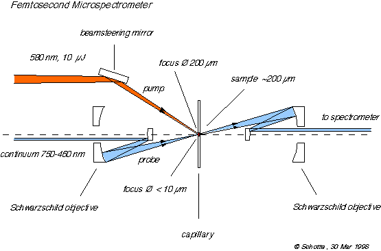
Sketch of setup

General Description:
A time-resolved absorption spectrometer has been setup up on the laser optical table. This instrument has been build for the preparation and as diagnostics of time-resolved X-ray diffraction experiments. It has been designed with single crystals of Myoglobin, Photoactive Yellow Protein or Bacteriorhodopsin in the order of 100 m m in mind, but there might be different applications in future as small molecule crystals or liquid jets.
It is important to have to following questions answered before attempting an X-ray diffraction experiment:
- What is the optimal pump wavelength to get a maximum percentage of excitation of your sample?
-
- How much pump energy and how many pulses can the sample tolerate without damage or showing irreversible changes?
-
- What is the degree of photolysis or excitation under the condition you would chose for the X-ray experiment.
-
A commonly used approach in laser spectroscopy is to send two time-delayed pulses through the sample, which originate from the same source and thus have a very reliable relative timing. The time delay is controlled by a mechanical delay line altering the time of flight of one pulse to the sample. Many spectrometers for femtosecond studies of proteins exist but they use beam sizes of a few millimeters and have been designed for liquid samples, large in volume and low in concentration compared to crystals. A focussing spectrometer especially suited for small single crystals has been designed by Janos Hajdu. However, it uses fiber optics and would not be compatible with fs pulses because of the pulse broadening in the fibers.
The spectrometer uses a small fraction of the 800 nm output of the Ti:Saphire amplifier to generate fs white light pulses by a process called continuum generation. The laser beam is focussed into 2 mm thick Sapphire crystal. Due to the high intensity at the focal point non-linear processes take over generate a smooth spectral distribution ranging from above 800 nm down to 460 nm.
Continuum generation is not completely understood yet. It is probably a combination of self phase modulation and stimulated Raman scattering. Self phase modulation is caused by the non-linearity of the refractive index. In the central part of the pulse where the intensity is highest the refractive is higher. Thus, the central part is retarded relative to the leading and trailing edge of the pulse. So the leading part is compressed and raised in frequency and the trailing part is expanded and lowered in frequency. Stimulated Raman scattering means that photon energy is partially transferred to high frequency lattice vibrations. Those, once excited, can then transfer energy back to other photons creating a blue-shifted continuum.
The non-linearity of the refractive index leads to self-focussing of the beam at the focal point. It contracts to a filament with a diameter of the order of the wavelength which does no diverge as long the intensity is high enough and the pulse is not stretched in length. A filament can contain a certain amount of energy. If the laser power is increased beyond a threshold a second filament will be formed, grow in intensity to a limit then a third one will appear and so on. A single-filament continuum is nearly a diffraction-limited source, which can be focussed to a very small sample area. A variable attenuator adjusts the incident laser power to give a maximum of intensity avoiding multi-filament formation. Sapphire crystal had been chosen because of its high damage threshold and the stability of the continuum. Continuum generation would work in glass as well but it is much easier to damage and even if the intensity is below the damage threshold it could locally alter the properties of the glass and make the continuum unstable or the source point move.
The continuum is imaged 1:1 to the sample by all-reflective optics so there is no chromatic aberration. The focussing optics is a "reflective microscope objective" made by Ealing. Although it is made of two spherical mirrors it has no spherical aberration. The ratio of the two focal lengths and their distance are chosen so the spherical aberration cancels out. Before the beam is sent to through the sample a beam splitter sends half of it directly to the spectrometer. This way transmission and a reference continuum spectrum can always be measured simultaneously. This is necessary because the unavoidable shot-to-shot fluctuations of the laser output not only alter the intensity but also the spectral distribution of the continuum. Both beams are focussed into a 200 m m optical fiber cable and guided to a spectrograph installed off the optical table. At this point the pulse broadening by the fiber is not a problem because it happens after the sample. The light is fed into the spectrograph by a multi-track fiber cable. It images the two fiber exits as separated traces onto a CCD chip.
There are computer-controlled shutters to block the pump or the probe beam selectively. The data acquisition program uses them to make automatically reference measurements to eliminate systematic errors. For each transient spectrum three additional measurements are made, pump beam blocked, probe beam blocked and both beams blocked.
This work has been done in collaboration with Prof. Philip Anfinrud from the Harvard University, starting in July 1997.
Characteristics
Probe beam size < 10 um Pump beam size 200 um Polarization linear or circular Spectral range 760-460 nm Spectral resolution 5 nm Time resolution 200 fs Time range 0-670 ps, n × 1.1 ms (n=1,2,…)


