- Home
- Users & Science
- Scientific Documentation
- ESRF Highlights
- ESRF Highlights 2014
- X-ray Imaging
- Robust lensless imaging with a high-flux instrument
Robust lensless imaging with a high-flux instrument
In the last decade, coherent diffractive imaging (CDI) has shown great promise as a dose-efficient and high-resolution complement to traditional (lens-based) X-ray microscopy. In contrast to conventional microscopy where an objective lensing system catches the diffracted X-ray photons to form a direct image on the detector, the X-ray photons are allowed to freely propagate to the detector forming a diffraction image. A computer algorithm is used to reconstruct an image of the specimen’s transmission from the traces left in such an indirect measurement. Ptychography is a CDI variant where the specimen is scanned transversally through the X-ray beam, collecting a diffraction image at each scan point [1,2]. Unlike traditional X-ray microscopy or other CDI techniques, ptychography can deliver quantitative amplitude and phase images of extended specimen beyond the resolution limit of lenses.
However, the benefits of diffraction imaging usually come at the cost of requiring “clean” diffraction data for the algorithms to work properly. Commonly this requirement is met by strong spectral and spatial filtering for beam purity in combination with a photon counting, pixel-array detector for an ideal measurement. While the first measure drastically reduces the available photon flux, the latter cannot distinguish single photon events at high flux. Ironically, this very need to reduce flux translates directly into lower achievable resolution [3].
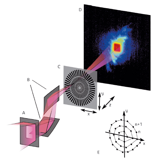
An experiment was carried out at the nano-imaging endstation of the former ID22 beamline (now ID16A) to investigate the robustness of ptychography in a configuration that allows for high-flux experiments, under conditions that were previously considered insufficient: a broad-bandwidth X-ray beam focused by a pair of crossed multilayer-coated Kirkpatrick-Baez mirrors and an integrating scintillator-based FReLoN detector (Figure 55). As can be seen in the diffraction image (D in Figure 55), the acquired diffraction data was degraded by a large halo around the central beam (bluish in the image). The high dynamic range in the diffraction signal caused this degradation by amplifying even faint effects like diffuse air scattering or a point spread in the scintillator coupled to the CCD.
 |
|
Fig. 55: Schematic of the experimental setup (not to scale) used for ptychography at the former ID22NI endstation. The X-ray beam enters through an aperture (A) and is focussed by two multilayer Kirkpatrick-Baez mirrors (B). The sample (C) is placed close to the focus and the diffraction patterns are recorded downstream by a CCD coupled to a scintillator (D). To avoid artifacts, the sample is scanned along a circular pattern (E). |
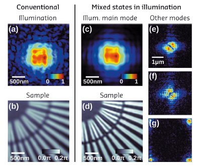
Consequently, conventional ptychographic algorithms struggled when reconstructing sample transmission (Figure 56b) and illuminating wavefront (Figure 56a) from these data. The illumination contains speckly noise and the phase image of the spoke-pattern of the sample is blurred.
Next, the mixed states approach [4] was applied, which is one of the latest algorithmic developments for the treatment of ptychographic data. It permits a number of concurrent modes to form the illumination and thereby emulates some deficiencies of the detector and the beam, e.g. partial coherence. These modifications alone allowed reconstruction of the sample in high detail and quality (Figure 56d) and the main mode (Figure 56c) now shows the shape expected for the illumination in the sample plane.
Furthermore, an analysis of the illumination modes revealed that the algorithm not only corrects for lower coherence and detector point spread (Figure 56, e and f), but also attempts to correct for an inhomogeneous response of the four detector quadrants in algorithmically less constrained parts of the illumination frame (Figure 56g).
 |
|
Fig. 56: Conventional reconstruction (left) compared with mixed-states approach (right). Conventional reconstruction: (a) modulus of the retrieved illumination wavefront (in a.u.) and (b) phase (in rad) of the reconstructed sample transmission function. Mixed state reconstruction: (c) showing the reconstruction of the main mode of the illumination and (d) phase of the sample transmission function. (e)-(g) an assortment of other modes reconstructed in the Illumination, where (e) and (f) can be associated with the detector point spread function or partial coherence while (g) is related to the inhomogeneous response of the four quadrants of the detector. |
Our work proves that mere adjustments in the data analysis steps can lead to dramatic improvements in image quality. With its newly demonstrated robustness, ptychography will be widely applicable with relaxed experimental requirements, paving the way to high-throughput, high-flux and high-resolution diffractive imaging.
Principal publication and authors
B. Enders (a), M. Dierolf (a), P. Cloetens (b), M. Stockmar (a), F. Pfeiffer (a) and P. Thibault (a,c), Appl. Phys. Lett. 104, 171104 (2014).
(a) Physics Department, Technische Universität München, Garching (Germany)
(b) ESRF
(c) Current address: Physics & Astronomy Department, University College London (UK)
References
[1] P. Thibault et al., Science 321, 379 (2008).
[2] M. Dierolf et al., Nature 467, 436 (2010).
[3] M. Howells et al., Journal of Electron Spectroscopy and Related Phenomena 170, 4 (2009).
[4] P. Thibault and A. Menzel, Nature 494, 68 (2013).


