- Home
- Users & Science
- Scientific Documentation
- ESRF Highlights
- ESRF Highlights 2015
- X-ray nanoprobe
- Residual strain in the mineral nanoparticles of human tooth dentine makes teeth stronger
Residual strain in the mineral nanoparticles of human tooth dentine makes teeth stronger
Human teeth operate in the mouth for many decades where they are subjected to huge forces and much wear and tear. The main tooth bulk material is dentine, which supports enamel externally, and is very tough and damage resistant. Evidence was found for the role of a previously overlooked toughening mechanism, arising from residual strain in the nanocomposite ultrastructure of dentine.
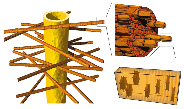
Dentine is a bone like biological nanocomposite, built of crystalline mineral particles, collagen fibres, and water [1]. This nanocomposite contains tubules, channels formed by odontoblast cells that produce dentine during tooth genesis. These cells typically reside in the pulp of normal ‘living’ teeth, sending out long ‘finger like’ extensions into the tubules. The tubules traverse the entire dentine tissue thickness, and they course through the mineralised collagen layers where they are surrounded by thick micrometre-sized ‘peritubular’ mineral sheaths (Figure 12). Mineralised collagen fibres are arranged in layers perpendicular to the tubules, and form a fibrous mass of inter-tubular dentine. Just how these different mineralised structures ‘work’ together to help dentine survive the excessive cyclic loading is still a mystery. This is particularly curious considering the lack of a self-healing biological mechanism such as remodelling, which is known to exist in bone.
 |
|
Fig. 12: Schematic representation of the dentine nanocomposite structure made of collagen fibres embedded in mineral, oriented orthogonally to dental tubules that lead to the pulp. The c-axis of the mineral particles in the fibres is aligned with the collagen axis. |
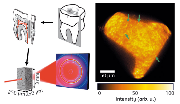
To shed additional light on the link between the micro-architecture of dentine and its remarkable structural integrity, we combined synchrotron-based diffraction methods at different length scales: a micrometre-sized beam in MySpot at BESSY, Germany and a nanofocused X-ray beam at the former ID22NI beamline (now ID16A) for diffraction nanotomography. We revealed important differences in the spatial arrangement of the nanometre-sized mineral crystallites. Diffraction nanotomography (see Figure 13) showed that crystallite alignment with the collagen fibres in the inter-tubular dentine differs from crystallite alignment in the peritubular dentine. The c-lattice parameters of apatite in differently treated samples of human tooth dentine were analysed along various azimuthal orientations of the radii of the apatite (002) Debye rings. The analysis, performed using XRDUA [2], showed that mineralised particles attached to dry collagen are under compression, and that compressive stresses vary as a function of humidity. Compression is no longer apparent when the collagen is destroyed by heating.
 |
|
Fig. 13: Slices cut from human teeth were used to create small pin-like bars which were mounted for diffraction-tomography at ID22NI. More than 15,000 diffraction images were analysed and the intensity distribution of the apatite 002 peak was mapped across the dentine microstructure. High intensity spots are distributed throughout the structure (green arrows) and correspond mainly to dentine tubules, with distinctly different mean orientation of the mineral particles, as compared to the general collagen-rich mineralised matrix, surrounding the tubules. |
We conclude that in the native tissue there is strong interaction of the mineral particles with the organic matrix that leads to significant compression of the mineral-particles, and we propose that this interaction is important for energy dissipation and damage resistance in teeth. This conclusion is corroborated by the observation that crystallites that are co-aligned with collagen fibres become more compressed than mineral particles of the tubules. The residual strains are completely eliminated by mild annealing at 250°C, i.e. when the organic component is destroyed [3]. The compressive strain and corresponding mineral-particle deformation correlate with the collagen layered arrangement, and presumably work against crack propagation from outside the tooth towards the pulp, across the layers, thus increasing resistance of the biostructure as a whole.
Engineers use internal stresses to strengthen materials for specific technical purposes, for example in concrete. Evolution has presumably ‘known’ about this trick for a long time, and has incorporated it into our natural teeth. Our findings suggest that residual stress and mineral compression mediated by the collagen/mineral interactions contribute significantly to the strengthening and toughening of teeth, complementing other toughening mechanisms and well-known structural strategies, acting in all similar bony biocomposites at larger length scales (e.g. microcracking). We believe that the balance of stresses between the nanoparticles and collagen and, especially, mineral compression initiated by the collagen/mineral bond, are important for the extended survival of teeth in the mouth.
Principal publication and authors
Compressive residual strains in mineral nanoparticles as a possible origin of enhanced crack resistance in human tooth dentin, J.-B. Forien (a), C. Fleck (b), P. Cloetens (c), G. Duda (a), P. Fratzl (d), E. Zolotoyabko (e) and P. Zaslansky (a), Nanoletters 15, 3729–3734 (2015); doi: 10.1021/acs.nanolett.5b00143.
(a) Julius Wolff Institute, Charité – Universitätsmedizin, Berlin (Germany)
(b) Materials Engineering, Berlin Institute of Technology, Berlin (Germany)
(c) ESRF
(d) Department of Biomaterials, Max-Planck-Institute of Colloids and Interfaces, Potsdam (Germany)
(e) Department of Materials Science and Engineering, Technion – Israel Institute of Technology, Haifa (Israel)
References
[1] P. Zaslansky, ‘Dentine’ in P. Fratzl (Ed.), Collagen: structure and mechanics, Springer, New York (2008), pp. 421–442.
[2] W. De Nolf et al., Appl. Crystallogr. 47, 1107–1117 (2014).
[3] S.R. Armstrong et al., J. Endod. 32, 638–641 (2006).


