- Home
- Users & Science
- Scientific Documentation
- ESRF Highlights
- ESRF Highlights 2015
- X-ray nanoprobe
- Analysis of solid oxide cell performance and degradation by X-ray phase nanotomography
Analysis of solid oxide cell performance and degradation by X-ray phase nanotomography
Efficiency and durability of solid oxide cells are important for the development of high temperature electrochemical converters. By analysing three-dimensional reconstructions of the electrodes obtained with X-ray phase nanotomography, a better understanding has been gained of the basic local phenomena that govern the macroscopic cell behaviour in terms of performance and degradation.
Solid oxide cells (SOCs) have potential for use as electrochemical converters operating at high temperatures. Their advantages include a high flexibility in terms of technological applications. For example, the same object can be alternatively used in fuel cell and steam electrolysis modes. In addition, high efficiency for hydrogen production and electrical power generation can be reached without the use of expensive electrocatalysts. Nevertheless, SOCs will become a competitive technology only if durability issues are solved.
SOCs consist of a ceramic multilayer cell, composed of a dense electrolyte sandwiched between two porous electrodes. The electrode microstructure plays a major role in the global electrolyser efficiency by controlling the rates of the electrochemical reactions. Moreover, evolution of the electrode microstructure in operation is widely correlated with cell degradation, even if the basic mechanisms are still not understood.
The global SOCs response is complex and depends on local electrode properties. Therefore, models describing the global SOC operation coupled to local visualisation of the fine electrode microstructure could help to unravel the SOCs operating mechanisms. An in-house multi-scale and multi-physics model has been developed at the CEA in collaboration with the ESRF [1-4]. Three dimensional reconstructions of electrodes are required in order to determine key model parameters for an accurate description of the electrode microstructure. To obtain both a large field of view and a high spatial resolution, X-ray phase nanotomography, or magnified holotomography, was selected for this study, using the former ESRF beamline ID22NI, now ID16A.
The methodology has been applied to different commercial cells composed of the typical SOCs materials. Hydrogen electrodes were made of a cermet of nickel and yttria stabilised zirconia (Ni-YSZ). The setup and data processing have been specifically adapted to handle such strongly absorbing ceramic materials by using harder X-rays and optimising the phase retrieval procedure. X-ray phase nanotomography was performed at an energy of 30 keV and electrode reconstructions were obtained with a field of view of 50 µm and a spatial resolution of about 75 nm (25 nm voxel size) [5].
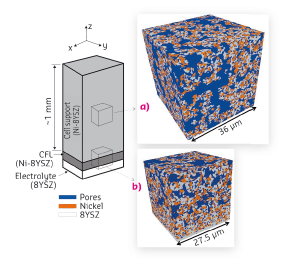
The reconstruction of a Ni-YSZ electrode bilayer (Figure 14) has been analysed numerically to compute all its morphological properties. Thanks to the large field of view of the reconstructions, all the microstructure properties have been successfully quantified with a high level of confidence [6,7]. These microstructural parameters were used to simulate the electrode overpotential at the microscopic scale as a function of temperature [6]. A deconvolution between the thermally activated processes has been proposed. This allows basic degradation mechanisms to be investigated.
 |
|
Fig. 14: 3D volume renderings of Ni-YSZ cermet obtained by magnified holotomography [5,6]. (a) Reconstruction taken in the cell support. (b) Reconstruction of the cathode functional layer (with a part of the support and the electrolyte). |
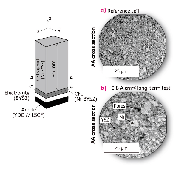
A major degradation phenomenon occurring in electrolysis mode corresponds to nickel agglomeration in the Ni-YSZ cathode. The Ni particle coarsening was observed by X-ray phase nanotomography. Microstructural properties extracted from the 3D reconstructions, such as the Ni/YSZ/gas triple phase boundary (TPB) lengths, exhibit a remarkable evolution during the long term tests (Figure 15). The evolution of morphological parameters was introduced in the micro and macro models to evaluate their impact on the cell response. It has been found that the decrease in density of TPB length results in a significant cell voltage increase, which explains the main part of the degradation rates.
 |
|
Fig. 15: Comparison of the microstructure of the Ni-YSZ electrode before and after operation showing nickel particle coarsening revealed by magnified X-ray holotomography. |
To enhance the predictive power of the simulation tools, physical models of Ni coarsening need to be calibrated. Therefore, a next step will be to obtain reconstructions of Ni particles before and after coarsening with improved spatial resolution at the new beamline ID16A.
Principal publication and authors
Degradation study by 3D reconstruction of a nickel–yttria stabilised zirconia cathode after high temperature steam electrolysis operation, E. Lay-Grindler (a), J. Laurencin (a), J. Villanova (b), P. Cloetens (b), P. Bleuet (a), A. Mansuy (a), J. Mougin (a) and G. Delette (a), Journal of Power Sources 269, 927-936 (2014); doi: 10.1016/j.jpowsour.2014.07.066.
(a) CEA-LITEN, Grenoble (France)
(b) ESRF
References
[1] J. Laurencin et al., J. Euro. Ceram. Soc. 28, 1857-1869 (2008).
[2] J. Laurencin et al., J. Power Sources 196, 2080–2093 (2011).
[3] G. Delette et al., Inter. J. Hydrogen Energy 38, 12379-12391 (2013).
[4] E. Lay-Grindler et al., Int. J. of Hydrogen Energy 38 6917-6929 (2013).
[5] J. Villanova et al., J. Power Sources 243, 841-849 (2013).
[6] F. Usseglio-Viretta et al., J. Power Sources 256, 394-403 (2014).
[7] J. Laurencin et al., J. Power Sources 198, 182-189 (2012).


