- Home
- News
- General News
- Cell telephony
Cell telephony
30-03-2012
ESRF users are cracking the structure of proteins that allow our cells to communicate, revealing important new drug targets.
Share
Do you rely on a shot of coffee to start the day? Thank G-protein coupled receptors (GPCRs) for your kick. These tiny proteins, which are embedded in cellular membranes, spring into action when molecules from inside or outside the body pass by, activating G-proteins inside the cell and triggering a physiological response. Some set off the retina’s reaction to light and the nose’s response to odours, while others bind to the hormones and neurotransmitters that regulate our moods or affect the strength of our heartbeat.
Over a quarter of all drugs on the market, including treatments for migraine, asthma and heart defects, act on the biochemical pathways stimulated by GPCRs. There are some 800 human GPCRs – half of which are devoted to detecting odours – but only a small proportion of the rest are targeted specifically by current drugs, leaving huge potential for researchers to find suitable activators or inhibitors for the treatment of a wide range of diseases.
Nobel aspirations
While researchers have understood much about the biochemical nature of GPCRs, little was known about how their structure influences the way that the receptor transmits signals through the cell membrane. That situation is changing rapidly thanks to synchrotrons like the APS and, in particular, the ESRF and its automated microfocus beamlines.
“The solution of GPCR structures is ripe for a Nobel prize,” says the ESRF’s Matt Bowler. “The first structure [of a GPCR that binds to hormones or neurotransmitters] solved four years ago involved a phenomenal amount of work after people had been banging their heads against the wall for over 15 years, but now the structures are coming thick and fast.”
The first structural solution of a human GPCR was rhodopsin, the receptor that responds to photons in our retinas, in 2000 – followed by a second structure in a different crystalline arrangement at the ESRF in 2004. But it was 2007 when researchers solved a GPCR that binds to hormones or neurotransmitters. Using the ESRF’s ID13 and ID23-2 microfocus beamlines, Brian Kobilka’s group at Stanford University in the US in collaboration with Gebhard Schertler and colleagues at the MRC Laboratory of Molecular Biology (LMB) in Cambridge, UK, determined the structure of the “beta-two adrenergic receptor” (β2AR), which is activated by adrenaline and lies behind the body’s “flight or fight” response by opening the airways in the lungs – making it an important target for β2AR agonists used in drugs to treat asthma.
“I have a very fond memory of the ESRF,” Kobilka told ESRFnews. “Gebhard brought us to the beamline in 2005 to test our first crystals and again in 2006 and 2007 to collect data that led to the first β2AR structure.”
The following year, in 2008, the LMB team used ID23-2 to determine the structure of the sister beta receptor, β1AR, which controls the speed of the heart. Knowledge of GPCR structures potentially allows drugs to be designed that bind specifically to only one subtype of receptor, reducing side effects that arise when drugs bind to other receptors.
 |
|
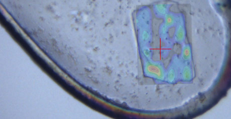
A G-coupled protein receptor crystal frozen in a nylon loop on beamline ID23-2, overlaid with a map showing the crystal’s diffraction quality. Credit: M Bowler. |
The struggle for structure
Unlike other proteins, such as haemoglobin solved 60 years ago, membrane proteins are only available in minute quantities and are unstable when removed from their cellular environment, which requires the use of detergents, and they continually undergo conformational “wobbles” even in the absence of an agonist. The key has been to find ways to purify receptors from the membrane so that they are locked in one particular conformation. One approach, developed by the LMB team, is to introduce thermo-stabilising mutations to the receptor, while US researchers have adopted a different approach by using “T4 lysozyme” fusions to reduce the receptor’s flexibility. “Fortunately, the two approaches give pretty much the same structure,” says Chris Tate, group leader at the LMB.
The ESRF’s microscopic beam size at ID23-2 has been vital to the recent successes. Structures can now be resolved at 2.5 Å resolution, showing in considerable atomic detail how ligands bind to the receptors.
The beta receptor structures determined in 2007 did not give up all of their secrets because they were all bound to antagonists and were therefore in an inactive state. Earlier this year, however, researchers determined how the beta receptors bind agonists, activating the receptors and relaying this signal to the G-protein inside the cell.
“The big breakthrough of 2011 has been a whole slew of receptor structures that are bound to agonists that initiate intracellular signalling,” says Tate, who was part of the team that cracked the β1AR’s antics at the ESRF after a six-month series of visits to ID23-2 last year. The real prize, says Tate, was the structure of the β2AR in complex with a G-protein determined by Kobilka and collaborators, which shows for the first time in molecular detail how receptors activate G-proteins.
Seven different GPCR structures have now been solved, with many more structures determined of receptors bound to different ligands. “With more than 350 GPCR structures left to solve, there is plenty of room for new players in the field,” says Tate. In addition to the all important beta receptors, the ESRF has helped the LMB team solve the structure of the adenosine A2A receptor, which among other things mediates the arousal effect of caffeine.
“The ESRF made an absolutely key contribution to all of these GPCR structures because it was the first [light source] to offer micro-crystallography, which then propagated to other sources,” says Schertler who is now at the Paul Scherrer Institute in Switzerland. “Pharmaceutical companies are starting to use this structural information already for drug-design strategies.”
The drive for structure determination of GPCRs has so far spun out three biotech companies – Heptares Therapeutics, Receptos and ConfometRx – and many other groups in both industry and academia are now engaged in the GPCR quest. There are parallels with the human genome project, says Bowler. “When people started decoding the genome, people thought it was madness because it would take too long and cost too much, but five years later two teams had developed the techniques and pulled it off.”
As for who might receive a telephone call from the Swedish Academy, ESRF users are sure to be among those in the running. But for Kobilka, who insists that many people lie behind the work he directs, the reward has already come. “There’s a lot of satisfaction in accomplishing such a major goal,” he told ESRFnews. “It is a special feeling to have.”
Nobel recognition for structural biology
 |
Winners of the 2009 Nobel Prize in Chemistry. Copyright © Nobel Media AB/Photo: Niclas Enberg. |
- 1946 Chemistry (J Sumner, half of prize): discovery that enzymes can be crystallised
- 1962 Physiology or Medicine (F Crick, J Watson and M Wilkins): molecular structure of nuclear acids and its significance for information transfer in living material
- 1962 Chemistry (M Perutz and J Kendrew): structures of globular proteins
- 1964 Chemistry (D Hodgkin): X-ray techniques to solve structures of important biochemical substances
- 1972 Chemistry (C Anfinsen, half of prize): studies of ribonuclease, especially concerning the connection between the amino acid sequence and the biologically active conformation
- 1982 Chemistry (A Klug): crystallographic electron microscopy and the structural elucidation of biologically important nucleic acid-protein complexes
- 1988 Chemistry (J Deisenhofer, R Huber and H Michel): 3D structure of a photosynthetic reaction centre
- 1997 Chemistry (P Boyer and J Walker, half of prize): mechanism underlying adenosine triphosphate synthesis
- 2002 Chemistry (J Fenn, K Tanaka and K Wüthrich): methods to identify and analyse structure of biological macromolecules
- 2003 Chemistry (P Agre and R MacKinnon): discoveries concerning channels in cell membranes
- 2006 Chemistry (R Kornberg): molecular basis of eukaryotic transcription
- 2009 Chemistry (V Ramakrishnan, T Steitz and A Yonath): structure and function of the ribosome
References
G Lebon et al. 2011 Nature 474 521–525.
R Moukhametzianov et al. 2011 PNAS 108 8228–8232.
S Rasmussen et al. 2007 Nature 450 383–387.
S Rasmussen et al. 2011 Nature 469 175–180.
C Riekel et al. 2005 Curr. Opin. Struct. Biol. 15 556–562.
D Rosenbaum et al. 2007 Science 318 1266–1273.
D Rosenbaum et al. 2011 Nature 469 236–240.
T Warne et al. 2008 Nature 454 486–491.
T Warne et al. 2011 Nature 469 241–244.
Matthew Chalmers
This article appeared in ESRFnews, October 2011.
To register for a free subscription and to rapidly receive the current issue, please go to: ESRFNews Digital Edition Subscription
Top image: Crystal structure of activated beta-2 adrenergic receptor β2AR (red) in complex with G-proteins (green, cyan and yellow). Credit: S.G.F. Rasmussen et al., Nature 477, 549–555 (2011).


