- Home
- News
- General News
- Human brain evolution,...
Human brain evolution, new insight through X-rays
07-09-2011
An experiment at the ESRF reveals the brain shape of an early human ancestor.
Share
A paper published today in Science reveals the highest resolution and most accurate X-ray scan ever made of the brain case of an early human ancestor. The insight derived from this data is like a powerful beacon on the hazy landscape of brain evolution across the transition from Australopithecus to Homo.
The publication is part of a series of five papers based on new evidence pertaining to various aspects of the anatomy of the species Australopithecus sediba (announced in April 2010 by Berger et al.) published in Science on 9 September 2011. Led by the University of the Witwatersrand in Johannesburg (South Africa), over 80 scientists from numerous institutes in Germany, the U.S., UK, Australia, France, South Africa and Switzerland worked on the project. The work on the brain included a scientist from the European Synchrotron Radiation Facility (ESRF) in Grenoble (France), where the X-ray microtomography scan was performed.
 |
|
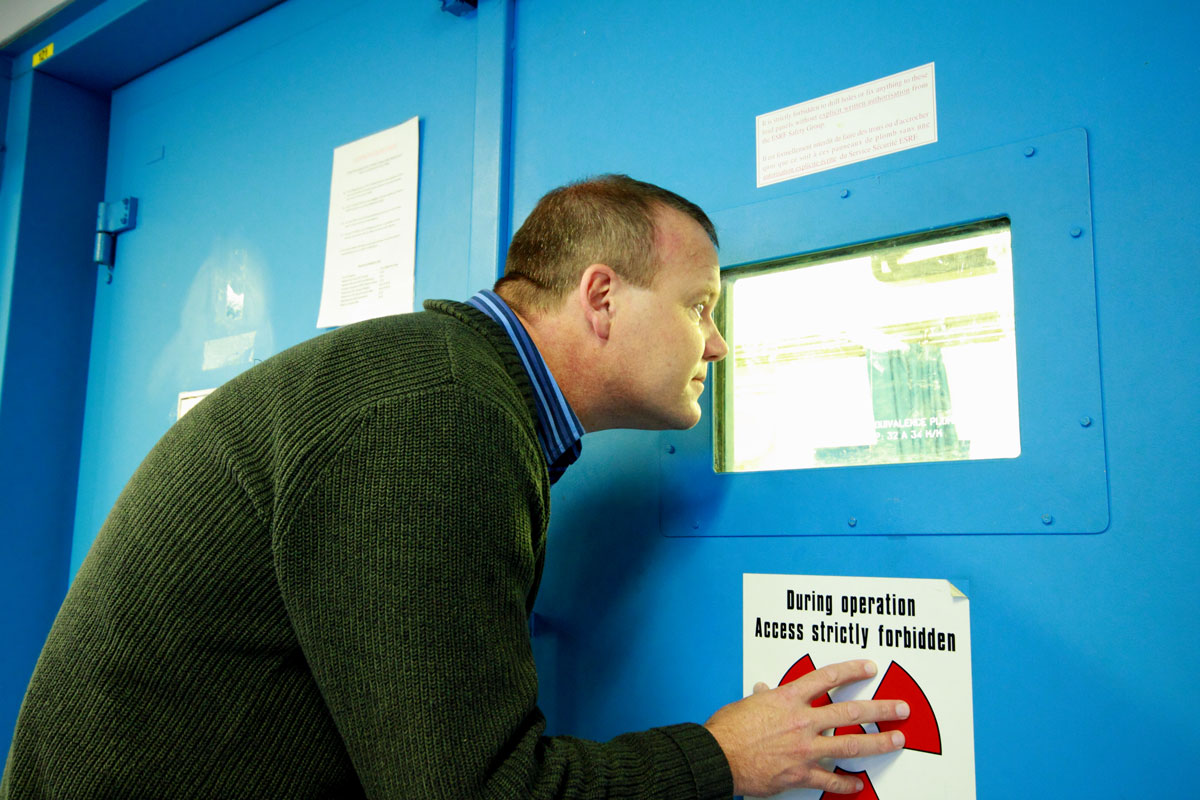
Lee Berger at the ESRF, looking through a glass window into beamline ID19 where the Australopithecus sediba is being scanned by X-rays. Credit ESRF/I. Montero. |
The exceptionally well-preserved cranium of MH 1 (Australopithecus sediba) was scanned at the ESRF at a resolution (3-D pixel size) of around 45 micrometres, just below the size of a human hair. Thanks to this high resolution, incredible details of the anatomy of sediba’s endocast could be revealed.
 |
|
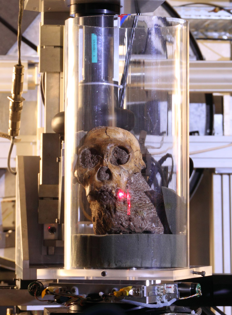
Close-up photo of the skull of Australopithecus sediba during the experiment at the ESRF beamline ID19. The red dot from a laser illustrates where the X-ray beam would meet the fossil. Credit ESRF/I. Montero. |
According to Prof. Lee Berger from the University of the Witwatersrand in Johannesburg (South Africa) who found the fossil in 2009, “the many very advanced features found in the brain and body make it possibly the best candidate ancestor for our genus, the genus Homo, more so than previous discoveries such as Homo habilis.”
Humans have a very large brain relative to their body size, about four times that of chimpanzees. Evolution from the brain of our shared ancestor with chimpanzees has seen this radical size increase. However, the reconstructed endocast (volume of the cranium) of MH1 is surprisingly small, with a volume of 420 cm3, on average only about 40 cm3 larger than chimpanzees.
 |
|
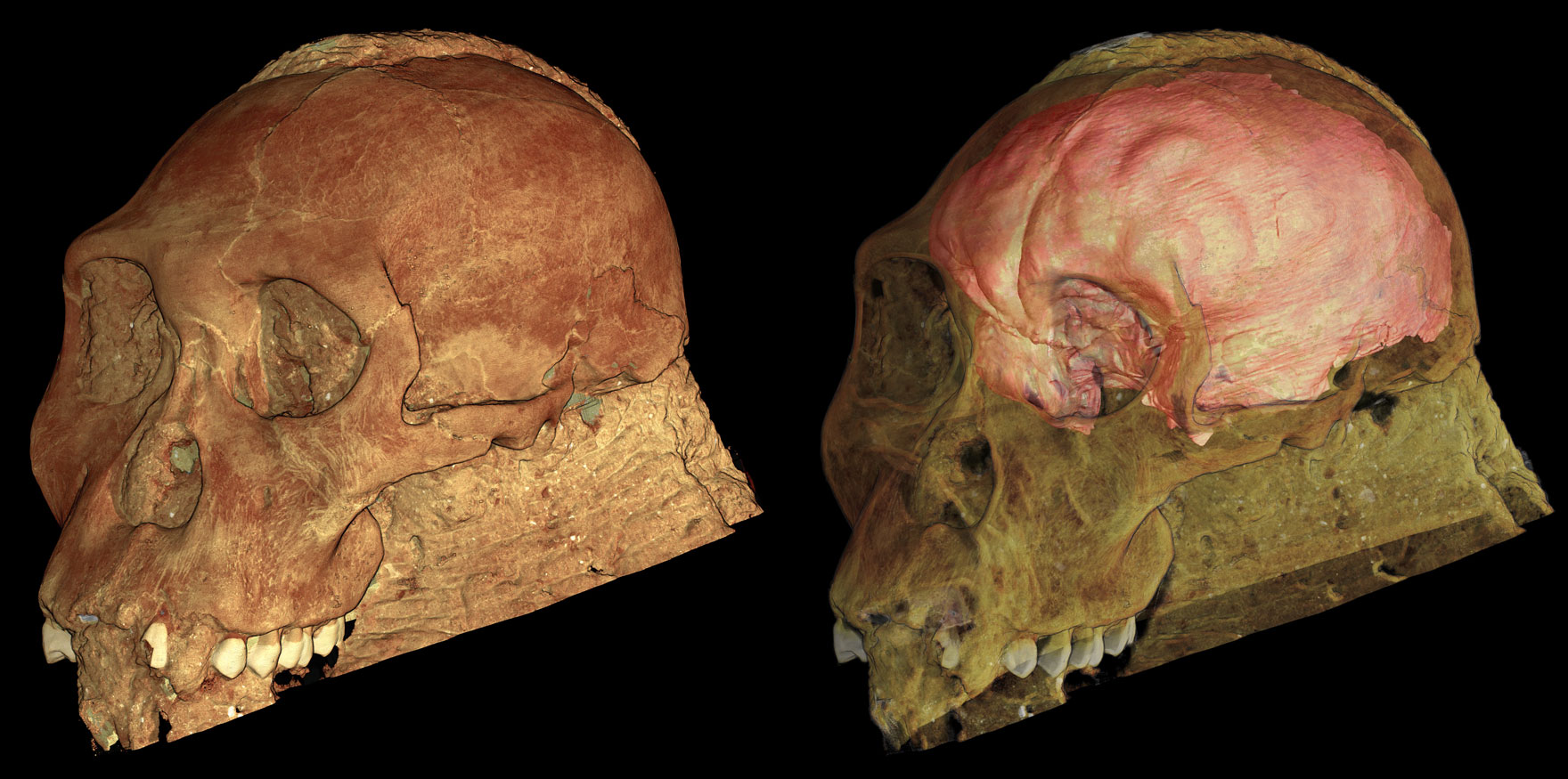
Reconstruction of the skull of Australopithecus sediba from an experiment at the ESRF beamline ID19. On the right, the skull is rendered partially transparent to show the brain endocast (depicted in pink). Courtesy of P. Tafforeau, ESRF. View high resolution image (1 mb). |
The study of this brain shows a surprising mix of characteristics. Its overall shape resembles humans more than chimpanzees and, given its small volume, this result is consistent with a model of gradual neural (brain) reorganisation in the front part of the brain. “Indeed, one of our major discoveries is that the shape and form of sediba’s brain is not consistent with a model of gradual brain enlargement, which has been hypothesised previously for the transition from Australopithecus to Homo”, adds Dr Kristian Carlson from the University of the Witwatersrand, who is the main author of the paper.
Use of synchrotron X-rays was instrumental for this discovery. The external shape of a brain is reflected, like in a mould, in the inner surface of a cranium. By mapping the contours of this internal surface, an image of the original brain located in the skull can therefore be produced. However, the skull of MH-1 was not emptied from bedrock after its discovery, and only the powerful X-rays at the ESRF could penetrate deep into the fossil to reveal the cranium’s interior shape at the desired resolution. Leaving the rock inside the cranium also ensured that its delicate inner surface was not damaged or altered during its extraction.
“The ESRF is the most powerful installation worldwide for scanning fossils, setting the standard for what can be achieved during non-destructive studies of internal structures of fossils,” concludes Paul Tafforeau, staff scientist at the ESRF and a co-author of the paper.
Reference
The Endocast of MH 1, Australopithecus sediba, K.J. Carlson, D. Stout, T. Jashashvili, D.J. de Ruiter, P. Tafforeau, K. Carlson, L.R. Berger, Science, 9 September 2011.
See also: Science Special Collection: Australopithecus sediba.
Top image: A 3-D rendering of the skull of Australopithecus sediba from an experiment at the ESRF beamline ID19. Credit ESRF/P. Tafforeau.
partners


European Synchrotron Radiation Facility - 71, avenue des Martyrs, CS 40220, 38043 Grenoble Cedex 9, France.